CD3 Recombinant Rabbit Monoclonal Antibody [JE80-02]

製品仕様
今すぐ注文,【现货】
問い合わせをクリックCatalog# HA720082
CD3 Recombinant Rabbit Monoclonal Antibody [JE80-02]
-
WB
-
IHC-P
-
IF-Tissue
-
IP
-
mIHC
-
IF-Cell
-
Human
-
unconjugated
概要
製品名
CD3 Recombinant Rabbit Monoclonal Antibody [JE80-02]
抗体の種類
Recombinant Rabbit monoclonal Antibody
免疫原
Synthetic peptide within human CD3E aa 158-207/207.
種属反応性
Human
検証された応用例
WB, IHC-P, IF-Tissue, IP, mIHC, IF-Cell
分子量
Predicted band size: 23 kDa
陽性対照
Jurkat cell lysates, human lymph nodes tissue, human spleen tissue, human small cell lung cancer, human non-small cell lung cancer, human gastric cancer.
結合
unconjugated
クローン番号
JE80-02
RRID
製品の特徴
形態
Liquid
保存方法
Shipped at 4℃. Store at +4℃ short term (1-2 weeks). It is recommended to aliquot into single-use upon delivery. Store at -20℃ long term.
保存用バッファー
PBS (pH7.4), 0.05% BSA, 40% Glycerol. Preservative: 0.05% Sodium Azide.
アイソタイプ
IgG
精製方法
Protein A affinity purified.
応用希釈度
-
WB
-
1:1,000-1:2,000
-
IF-Cell
-
1:100
-
IHC-P
-
1:600-1:1,000
-
IF-Tissue
-
1:100-1:200
-
mIHC
-
1:500-1:1,500
-
IP
-
Use at an assay dependent concentration.
論文における応用例
| mIHC | 確認する 5 以下の論文 |
| IHC-P | 確認する 3 以下の論文 |
| IF | 確認する 2 以下の論文 |
| IHC | 確認する 2 以下の論文 |
| IF-Tissue | 確認する 1 以下の論文 |
論文における種属
| Human | 確認する 8 以下の論文 |
| Mouse | 確認する 6 以下の論文 |
| Rat | 確認する 1 以下の論文 |
ターゲット
機能
The CD3 protein is a T-cell marker, a complex of four structurally distinct membrane glycoprotein isoforms, 20-50 kDa, comprising extracellular, transmembrane and intracellular domains. The CD3 complex is responsible for mediating signal transduction to the internal environment upon antigenic recognition by TCR, causing T-cell proliferation and release of cytokines. Except for a weak expression in Purkinje cells (with some of the Abs) and activated NK-cells, CD3 is found only in T-cells. CD3 appear in the cytoplasm of prothymocytes, and on the surface of about 95% of thymocytes, while cytoplasmic CD3 is lost as the cells differentiate into medullary thymocytes. In therapy resistant celiac disease, a shift from membranous to cytoplasmic CD3 expression is seen (together with loss of CD8). In malignant lymphomas, CD3 is a pan-T-cell lineage-restricted antigen, detected in 80-97% of the T-cell lymphomas. Mature T-cell lymphomas including cases of mycosis fungoides, peripheral T-cell lymphoma and anaplastic large cell lymphoma may aberrantly lose CD3. NK-cell lymphomas can show a cytoplasmic reaction. Reed-Sternberg cells may show a globular paranuclear reaction. CD3 is an important marker in the classification of malignant lymphomas and lymphoid leukaemias. Also the marker is useful for the identification of T-cells in, e.g., celiac disease, lymphocytic colitis and colorectal carcinomas associated with loss of a mismatch repair protein.
背景文献
1. Erman B. et. al. Biallelic Form of a Known CD3E Mutation in a Patient with Severe Combined Immunodeficiency. J Clin Immunol. 2020 Apr
2. Chen Q. et. al. CD3(+)CD20(+) T cells and their roles in human diseases. Hum Immunol. 2019 Mar
亜細胞局在
Cell membrane.
UNIPROT #
別名
4930549J05Rik antibody
A430104F18Rik antibody
AW552088 antibody
Cd247 antibody
CD247 antigen antibody
CD247 antigen, zeta subunit antibody
CD247 molecule antibody
CD3 antibody
CD3 antigen, delta subunit antibody
CD3 delta antibody
展開4930549J05Rik antibody
A430104F18Rik antibody
AW552088 antibody
Cd247 antibody
CD247 antigen antibody
CD247 antigen, zeta subunit antibody
CD247 molecule antibody
CD3 antibody
CD3 antigen, delta subunit antibody
CD3 delta antibody
CD3 epsilon antibody
CD3 eta antibody
CD3 gamma antibody
CD3 molecule delta polypeptide antibody
CD3 molecule, epsilon polypeptide antibody
CD3 molecule, gamma polypeptide antibody
CD3 zeta antibody
CD3-DELTA antibody
CD3d antibody
CD3D antigen delta polypeptide antibody
CD3d antigen, delta polypeptide (TiT3 complex) antibody
CD3d molecule delta antibody
CD3d molecule delta CD3 TCR complex antibody
CD3d molecule, delta (CD3-TCR complex) antibody
CD3D_HUMAN antibody
CD3E antibody
CD3e antigen antibody
CD3E antigen epsilon polypeptide antibody
CD3e antigen, epsilon polypeptide (TiT3 complex) antibody
CD3e molecule epsilon antibody
CD3e molecule epsilon CD3 TCR complex antibody
CD3e molecule, epsilon (CD3-TCR complex) antibody
CD3epsilon antibody
CD3G antibody
CD3g antigen antibody
CD3G antigen gamma polypeptide antibody
CD3g antigen, gamma polypeptide (TiT3 complex) antibody
CD3g molecule gamma antibody
CD3g molecule gamma CD3 TCR complex antibody
CD3g molecule, gamma (CD3-TCR complex) antibody
CD3H antibody
CD3Q antibody
CD3Z antibody
CD3zeta antibody
Ctg3 antibody
FLJ17620 antibody
FLJ17664 antibody
FLJ18683 antibody
FLJ79544 antibody
FLJ94613 antibody
IMD19 antibody
Leu-4 antibody
MGC138597 antibody
OKT3, delta chain antibody
OTTHUMP00000032544 antibody
T cell receptor antibody
T cell receptor T3 delta chain antibody
T cell receptor T3 gamma chain antibody
T cell receptor T3 zeta chain antibody
T cell receptor zeta chain antibody
T cell surface antigen T3/Leu 4 epsilon chain antibody
T cell surface glycoprotein CD3 antibody
T cell surface glycoprotein CD3 delta chain antibody
T cell surface glycoprotein CD3 epsilon chain antibody
T cell surface glycoprotein CD3 gamma chain antibody
T cell surface glycoprotein CD3 zeta chain antibody
T-cell antigen receptor complex, delta subunit of T3 antibody
T-cell antigen receptor complex, epsilon subunit of T3 antibody
T-cell antigen receptor complex, gamma subunit of T3 antibody
T-cell antigen receptor complex, zeta subunit of CD3 antibody
T-cell receptor T3 delta chain antibody
T-cell receptor T3 gamma chain antibody
T-cell surface antigen T3/Leu-4 epsilon chain antibody
T-cell surface glycoprotein CD3 delta chain antibody
T-cell surface glycoprotein CD3 epsilon chain antibody
T-cell surface glycoprotein CD3 gamma chain antibody
T3 antibody
T3d antibody
T3e antibody
T3g antibody
T3z antibody
TCRE antibody
TCRk antibody
Tcrz antibody
TCRzeta antibody
折りたたむ画像
-

Western blot analysis of CD3 on Jurkat cell lysates with Rabbit anti-CD3 antibody (HA720082) at 0.5μg/ml dilution.
Lysates/proteins at 10 µg/Lane.
Predicted band size: 23 kDa
Observed band size: 23 kDa
Exposure time: 2 minutes;
15% SDS-PAGE gel.
Proteins were transferred to a PVDF membrane and blocked with 5% NFDM/TBST for 1 hour at room temperature. The primary antibody (HA720082) at 0.5μg/ml dilution was used in 5% NFDM/TBST at room temperature for 2 hours. Goat Anti-Rabbit IgG - HRP Secondary Antibody (HA1001) at 1:200,000 dilution was used for 1 hour at room temperature. -

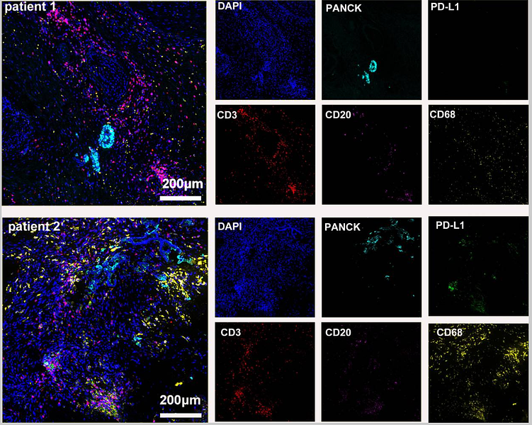
Fluorescence multiplex immunohistochemical analysis of Tertiary Lymphoid Structures in Human Small Cell Lung Cancer (Formalin/PFA-fixed paraffin-embedded sections). Panel A: the merged image of anti-CD20 (HA721138, green), anti-PD-L1 (HA721176, cyan), anti-CD56 (ET1702-43, magenta) and anti-CD3 (HA720082, yellow) on tertiary lymphoid structures. Panel B: anti- CD20 stained on B cells. Panel C: anti-PD-L1 stained on dendritic cells and macrophages cells. Panel D: anti-CD56 stained on NKT cells. Panel E: anti-CD3 stained on T cells. HRP Conjugated UltraPolymer Goat Polyclonal Antibody HA1119/HA1120 was used as a secondary antibody. The immunostaining was performed with the Sequential Immuno-staining Kit (IRISKit™MH010101, www.luminiris.cn). The section was incubated in four rounds of staining: in the order of HA721138 (1/1,500 dilution), HA721176 (1/1,000 dilution), ET1702-43 (1/1,000 dilution), and HA720082 (1/500 dilution) for 20 mins at room temperature. Each round was followed by a separate fluorescent tyramide signal amplification system. Heat mediated antigen retrieval with Tris-EDTA buffer (pH 9.0) for 30 mins at 95℃. DAPI (blue) was used as a nuclear counter stain. Image acquisition was performed with Olympus VS200 Slide Scanner.
-

Fluorescence multiplex immunohistochemical analysis of the human non-small cell lung cancer (Formalin/PFA-fixed paraffin-embedded sections). Panel A: the merged image of anti-CD20 (HA721138, green), anti-CD68 (HA601115, gray), anti-PD-L1 (HA721176, cyan), anti-panCK (HA601138, magenta) and anti-CD3 (HA720082, yellow) on human non-small cell lung cancer. Panel B: anti- CD20 stained on B cells. Panel C: anti-CD68 stained on macrophage M1 and macrophage M2. Panel D: anti-PD-L1 stained on dendritic cells and macrophages cells. Panel E: anti-panCK stained on cancer cells. Panel F: anti-CD3 stained on T cells. HRP Conjugated UltraPolymer Goat Polyclonal Antibody HA1119/HA1120 was used as a secondary antibody. The immunostaining was performed with the Sequential Immuno-staining Kit (IRISKit™MH010101, www.luminiris.cn). The section was incubated in five rounds of staining: in the order of HA721138 (1/1,500 dilution), HA601115 (1/2,000 dilution), HA721176 (1/1,000 dilution), HA601138 (1/3,000 dilution), and HA720082 (1/500 dilution) for 20 mins at room temperature. Each round was followed by a separate fluorescent tyramide signal amplification system. Heat mediated antigen retrieval with Tris-EDTA buffer (pH 9.0) for 30 mins at 95℃. DAPI (blue) was used as a nuclear counter stain. Image acquisition was performed with Olympus VS200 Slide Scanner.
-

Fluorescence multiplex immunohistochemical analysis of the human gastric cancer (Formalin/PFA-fixed paraffin-embedded sections). Panel A: the merged image of anti-CD31 (M1511-8, red), anti-αSMA (ET1607-53, gray), anti-CD11b (ET1706-04, cyan), anti-panCK (HA601138, magenta) and anti-CD3 (HA720082, yellow) on human gastric cancer. Panel B: anti- CD31 stained on the endothelial cells. Panel C: anti-αSMA stained on cancer-associated fibroblasts and smooth muscle cells. Panel D: anti-CD11b stained on myeloid cells. Panel E: anti-panCK stained on cancer cells. Panel F: anti-CD3 stained on T cells. HRP Conjugated UltraPolymer Goat Polyclonal Antibody HA1119/HA1120 was used as a secondary antibody. The immunostaining was performed with the Sequential Immuno-staining Kit (IRISKit™MH010101, www.luminiris.cn). The section was incubated in five rounds of staining: in the order of M1511-8 (1/1,000 dilution), ET1607-53 (1/2,000 dilution), ET1706-04 (1/1,000 dilution), HA601138 (1/3,000 dilution), and HA720082 (1/500 dilution) for 20 mins at room temperature. Each round was followed by a separate fluorescent tyramide signal amplification system. Heat mediated antigen retrieval with Tris-EDTA buffer (pH 9.0) for 30 mins at 95℃. DAPI (blue) was used as a nuclear counter stain. Image acquisition was performed with Olympus VS200 Slide Scanner.
-

Fluorescence multiplex immunohistochemical analysis of the human gastric cancer (Formalin/PFA-fixed paraffin-embedded sections). Panel A: the merged image of anti-Ki67 (HA721115, red), anti-CD31 (M1511-8, green), anti-CD3 (HA720082, cyan), anti-panCK (HA601138, magenta) and anti-αSMA (ET1607-53, yellow) on human gastric cancer. Panel B: anti- Ki67 stained on cells in G1, S, G2 and M phases of cell cycle. Panel C: anti-CD31 stained on the endothelial cells. Panel D: anti-CD3 stained on T cells. Panel E: anti-panCK stained on cancer cells. Panel F: anti-αSMA stained on cancer-associated fibroblasts and smooth muscle cells. HRP Conjugated UltraPolymer Goat Polyclonal Antibody HA1119/HA1120 was used as a secondary antibody. The immunostaining was performed with the Sequential Immuno-staining Kit (IRISKit™MH010101, www.luminiris.cn). The section was incubated in five rounds of staining: in the order of HA721115 (1/2,000 dilution), M1511-8 (1/1,000 dilution), HA720082 (1/500 dilution), HA601138 (1/3,000 dilution), and ET1607-53 (1/2,000 dilution) for 20 mins at room temperature. Each round was followed by a separate fluorescent tyramide signal amplification system. Heat mediated antigen retrieval with Tris-EDTA buffer (pH 9.0) for 30 mins at 95℃. DAPI (blue) was used as a nuclear counter stain. Image acquisition was performed with Olympus VS200 Slide Scanner.
-

Fluorescence multiplex immunohistochemical analysis of human gastric cancer (Formalin/PFA-fixed paraffin-embedded sections). Panel A: the merged image of anti-CD11b (ET1706-04, Red), anti-CD3 (HA720082, Green) and anti-CD31 (M1511-8, Yellow) on human gastric cancer. HRP Conjugated UltraPolymer Goat Polyclonal Antibody HA1119/HA1120 was used as a secondary antibody. The immunostaining was performed with the Sequential Immuno-staining Kit (IRISKit™MH010101, www.luminiris.cn). The section was incubated in three rounds of staining: in the order of ET1706-04 (1/1,000 dilution), HA720082 (1/500 dilution) and M1511-8 (1/1,000 dilution) for 20 mins at room temperature. Each round was followed by a separate fluorescent tyramide signal amplification system. Heat mediated antigen retrieval with Tris-EDTA buffer (pH 9.0) for 30 mins at 95℃. DAPI (blue) was used as a nuclear counter stain. Image acquisition was performed with Zeiss Observer 7 Inverted Fluorescence Microscope.
-

Immunohistochemical analysis of paraffin-embedded human lymph nodes tissue with Rabbit anti-CD3 antibody (HA720082) at 1/600 dilution.
The section was pre-treated using heat mediated antigen retrieval with Tris-EDTA buffer (pH 9.0) for 20 minutes. The tissues were blocked in 1% BSA for 20 minutes at room temperature, washed with ddH2O and PBS, and then probed with the primary antibody (HA720082) at 1/600 dilution for 1 hour at room temperature. The detection was performed using an HRP conjugated compact polymer system. DAB was used as the chromogen. Tissues were counterstained with hematoxylin and mounted with DPX. -

Immunohistochemical analysis of paraffin-embedded human spleen tissue with Rabbit anti-CD3 antibody (HA720082) at 1/1,000 dilution.
The section was pre-treated using heat mediated antigen retrieval with Tris-EDTA buffer (pH 9.0) for 20 minutes. The tissues were blocked in 1% BSA for 20 minutes at room temperature, washed with ddH2O and PBS, and then probed with the primary antibody (HA720082) at 1/1,000 dilution for 1 hour at room temperature. The detection was performed using an HRP conjugated compact polymer system. DAB was used as the chromogen. Tissues were counterstained with hematoxylin and mounted with DPX. -

Immunocytochemistry analysis of Jurkat cells labeling CD3 with Rabbit anti-CD3 antibody (HA720082) at 1/100 dilution.
Cells were fixed in 4% paraformaldehyde for 20 minutes at room temperature, permeabilized with 0.1% Triton X-100 in PBS for 5 minutes at room temperature, then blocked with 1% BSA in 10% negative goat serum for 1 hour at room temperature. Cells were then incubated with Rabbit anti-CD3 antibody (HA720082) at 1/100 dilution in 1% BSA in PBST overnight at 4 ℃. Goat Anti-Rabbit IgG H&L (iFluor™ 488, HA1121) was used as the secondary antibody at 1/1,000 dilution. PBS instead of the primary antibody was used as the secondary antibody only control. Nuclear DNA was labelled in blue with DAPI.
Beta tubulin (M1305-2, red) was stained at 1/100 dilution overnight at +4℃. Goat Anti-Mouse IgG H&L (iFluor™ 594, HA1126) was used as the secondary antibody at 1/1,000 dilution. -

Immunofluorescence analysis of paraffin-embedded human lymph nodes tissue labeling CD3 with Rabbit anti-CD3 antibody (HA720082) at 1/100 dilution.
The section was pre-treated using heat mediated antigen retrieval with Tris-EDTA buffer (pH 9.0) for 20 minutes. The tissues were blocked in 10% negative goat serum for 1 hour at room temperature, washed with PBS, and then probed with the primary antibody (HA720082, green) at 1/100 dilution overnight at 4 ℃, washed with PBS.
Goat Anti-Rabbit IgG H&L (iFluor™ 488, HA1121) was used as the secondary antibody at 1/1,000 dilution. Nuclei were counterstained with DAPI (blue). -

CD3 was immunoprecipitated from 1mg/ml Jurkat whole cell lysate with HA720082 at 2ug/ml dilution. Western blot was performed from the immunoprecipitate using HA720082 at 1ug/ml dilution. Goat anti-Rabbit IgG-HRP antibody (HA1001) was used as the secondary antibody at 1/300,000 dilution.
Lane 1: Jurkat whole cell lysate 5 μg (Input).
Lane 2: HA720082 IP in Jurkat whole cell lysate.
Lane 3: Rabbit monoclonal IgG instead of HA720082 in Jurkat whole cell lysate.
Blocking and dilution buffer and concentration: 5% NFDM/TBST.
Exposure time: 30 seconds. -

Application: Immunohistochemistry (IHC-P)
Species: Human
Tissue: Appendix
Sample: Paraffin-embedded section
Primary antibody dilution: 1/1,000
Antigen retrieval: ER2
Platform: Leica Biosystems BOND® RX -

Application: Immunohistochemistry (IHC-P)
Species: Human
Tissue: Appendix
Sample: Paraffin-embedded section
Primary antibody dilution: 1/1,000
Antigen retrieval: ER2
Platform: Leica Biosystems BOND® RX
ご注意ください: All products are "FOR RESEARCH USE ONLY AND ARE NOT INTENDED FOR DIAGNOSTIC OR THERAPEUTIC USE"
論文での実績
-
Epithelial pyroptosis-induced TREM1+ macrophages activate Th17 cells to accelerate oral mucosal inflammation
Author: Shang Qianhui, Wang Ziyuan, Peng Jiakuan, Yang Dan, Li Weiqi, Huang Xiaoyu, Qing Maofeng, Cheng Hao, Liu Jiaxin, Dan Hongxia, Zeng Xin, Zhou Yu, Zhang Dunfang, Xu Hao, Chen Qianming
PMID: 10.1038/s41420-025-02853-7
Journal: Cell Death Discovery
アプリケーション: mIHC
交差性: Human
掲載日: 2025 Nov
-
Citation
-
Harnessing NKG2D CAR-T cells with radiotherapy: a novel approach for esophageal squamous cell carcinoma treatment
Author: Tianyu Liu, Liyuan Fan, Weicheng Huang, Pengxiang Chen, Yuchen Liu, Shuyun Wang, Kaiyue Guo, Yufeng Cheng, Yali Han
PMID: 40510363
Journal: Frontiers In Immunology
アプリケーション: IHC
交差性: Mouse
掲載日: 2025 May
-
Citation
-
Decellularized lymph node scaffolds accelerate restoration of lymphatic drainage in rat hind limb lymphedema
Author: Yang Jian, Jian Zhou, Wenjie Pan, Jiayin Chen, Yanji Zhang, Yanqi Li, Xin Liu, Shune Xiao, Chenliang Deng, Zairong Wei
PMID: 10.1002/btm2.70056
Journal: Bioengineering & Translational Medicine
アプリケーション: mIHC,IHC
交差性: Rat,Mouse
掲載日: 2025 Jul
-
Citation
-
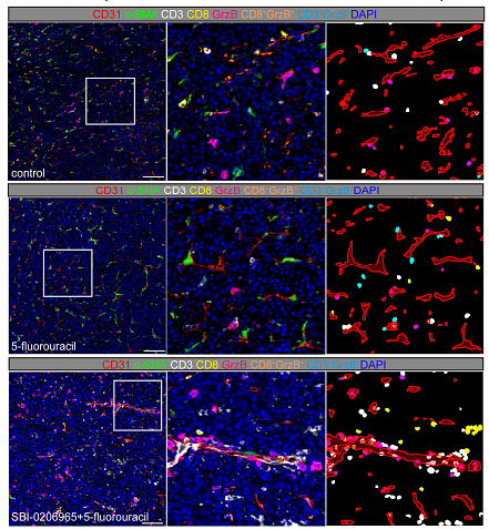
Inhibiting autophagy selectively prunes dysfunctional tumor vessels and optimizes the tumor immune microenvironment
Author:
PMID: 10.7150/thno.98285
Journal: Theranostics
アプリケーション: mIHC
交差性: Mouse
掲載日: 2025 Jan
-
Citation
-
Systematic evaluation of intratumoral and peripheral BCR repertoires in three cancers
Author: Sofia V Krasik, Ekaterina A Bryushkova, George V Sharonov, Daria S Myalik, Elizaveta V Shurganova, Dmitry V Komarov, Irina A Shagina, Polina S Shpudeiko, Maria A Turchaninova, Maria T Vakhitova, Igor V Samoylenko, Dimitr T Marinov, Lev V Demidov, Vladimir E Zagaynov, Dmitriy M Chudakov, Ekaterina O Serebrovskaya
PMID: 39831798
Journal: eLife
アプリケーション: IF
交差性: Human
掲載日: 2025 Jan
-
Citation
-
Inhibiting autophagy selectively prunes dysfunctional tumor vessels and optimizes the tumor immune microenvironment
Author: Wanting Hou, Chaoxin Xiao, Ruihan Zhou, Xiaohong Yao, Qin Chen, Tongtong Xu, Fujun Cao, Yulin Wang, Xiaoying Li, Ouying Yan, Xiaolin Ai, Cheng Yi, Dan Cao, Chengjian Zhao
PMID: 39744218

Journal: Theranostics
アプリケーション: mIHC
交差性: Mouse
掲載日: 2025 Jan
-
Citation
-
Systematic evaluation of intratumoral and peripheral BCR repertoires in three cancers
Author:
PMID: 10.7554/eLife.89506
Journal: eLife
アプリケーション: IF-Tissue
交差性: Human
掲載日: 2025 Jan
-
Citation
-
Targeting LHPP in neoadjuvant chemotherapy resistance of gastric cancer: insights from single-cell and multi-omics data on tumor immune microenvironment and stemness characteristics
Author: Gao You-Xin, Guo Xiao-Jing, Lin Bin, Huang Xiao-Bo, Tu Ru-Hong, Lin Mi, Cao Long-Long, Chen Qi-Yue, Wang Jia-Bin, Xie Jian-Wei, Li Ping, Zheng Chao-Hui, Yang Ying-Hong, Huang Chang-Ming, Lin Jian-Xian
PMID: 40240758
Journal: Cell Death & Disease
アプリケーション: IHC-P
交差性: Human,Mouse
掲載日: 2025 Apr
-
Citation
-
A humanized mouse model to study asthmatic airway remodeling and Muc-5ac secretion via the human IL-33
Author: Zhang Dong,et al
PMID: 38226717
Journal: Allergy Asthma & Immunology Research
アプリケーション: IHC-P
交差性: Human,Mouse
掲載日: 2024 May
-
Citation
-
Case report: Diverse immune responses in advanced pancreatic ductal adenocarcinoma treated with immune checkpoint inhibitor-based conversion therapies
Author: Li Xiaoying, Xiao Chaoxin, Li Ruizhen, Zhang Pei, Cao Dan
PMID: 38415262

Journal: Frontiers In Immunology
アプリケーション: mIHC
交差性: Human
掲載日: 2024 Feb
-
Citation
-
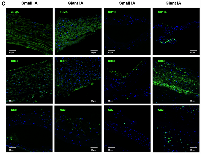
Decoding the Cell Atlas and Inflammatory Features of Human Intracranial Aneurysm Wall by Single‐Cell RNA Sequencing
Author: Hang Ji, Yue Li, Haogeng Sun, Ruiqi Chen, Ran Zhou, Yongbo Yang, Rong Wang, Chao You, Anqi Xiao, Liu Yi
PMID: 38390814

Journal: Journal Of The American Heart Association
アプリケーション: IF
交差性: Human
掲載日: 2024 Feb
-
Citation
-
Spatial patterns and MRI-based radiomic prediction of high peritumoral tertiary lymphoid structure density in hepatocellular carcinoma: a multicenter study
Author: Shichao Long,et al
PMID: 39675785
Journal: Journal For Immunotherapy Of Cancer
アプリケーション: IHC-P
交差性: Human
掲載日: 2024 Dec
-
Citation
同じターゲット & 同じ経路の製品
CD3 Recombinant Rabbit Monoclonal Antibody
Application: mIHC
Reactivity: Human
Conjugate: unconjugated
CD3 Recombinant Rabbit Monoclonal Antibody [PSH19-70] - BSA and Azide free
Application: WB,IHC-P,IF-Cell,FC,mIHC
Reactivity: Human,Mouse,Rat
Conjugate: unconjugated
CD3 Recombinant Rabbit Monoclonal Antibody [PSH19-70]
Application: WB,IHC-P,IF-Cell,FC,mIHC
Reactivity: Human,Mouse,Rat
Conjugate: unconjugated
CD3 Recombinant Mouse Monoclonal Antibody [PD01-43]
Application: WB,IHC-P,IF-Tissue
Reactivity: Human
Conjugate: unconjugated


