Glucose Transporter GLUT1 Recombinant Rabbit Monoclonal Antibody [SA0377]

製品仕様
今すぐ注文,【现货】
問い合わせをクリックCatalog# ET1601-10
Glucose Transporter GLUT1 Recombinant Rabbit Monoclonal Antibody [SA0377]
-
WB
-
IF-Cell
-
IF-Tissue
-
IHC-P
-
FC
-
mIHC
-
IHC-Fr
-
Human
-
Mouse
-
Rat
-
HA750002
不含抗保成分
-
ET1601-10
含抗保成分
-
unconjugated
概要
製品名
Glucose Transporter GLUT1 Recombinant Rabbit Monoclonal Antibody [SA0377]
抗体の種類
Recombinant Rabbit monoclonal Antibody
免疫原
Synthetic peptide within Human GLUT1 aa 443-492 / 492.
種属反応性
Human, Mouse, Rat
検証された応用例
WB, IF-Cell, IF-Tissue, IHC-P, FC, mIHC, IHC-Fr
分子量
Predicted band size: 54 kDa
陽性対照
HeLa cell lysate, PC-12 cell lysate, HeLa, HT-29 cell lysate, HepG2 cell lysate, NIH/3T3 cell lysate, L-929 cell lysate, mouse brain tissue lysate, rat brain tissue lysate, Jurkat, NIH/3T3, C6, human liver tissue, human placenta tissue, human liver carcinoma tissue, human kidney tissue, mouse liver tissue, mouse kidney tissue, HepG2, human lung cancer tissue, human liver tissue.
結合
unconjugated
クローン番号
SA0377
RRID
製品の特徴
形態
Liquid
保存方法
Shipped at 4℃. Store at +4℃ short term (1-2 weeks). It is recommended to aliquot into single-use upon delivery. Store at -20℃ long term.
保存用バッファー
1*TBS (pH7.4), 0.05% BSA, 40% Glycerol. Preservative: 0.05% Sodium Azide.
アイソタイプ
IgG
精製方法
Protein A affinity purified.
応用希釈度
-
WB
-
1:50,000-1:100,000
-
IF-Cell
-
1:500-1:1,000
-
IHC-P
-
1:5,000-1:10,000
-
IF-Tissue
-
1:500-1:1,000
-
FC
-
1:500-1:1,000
-
mIHC
-
1:30,000
-
IHC-Fr
-
1:1,000-1:2,000
論文における応用例
| WB | 確認する 14 以下の論文 |
| IF | 確認する 6 以下の論文 |
| IF-tissue | 確認する 1 以下の論文 |
| IF-Cell | 確認する 1 以下の論文 |
| IHC-P | 確認する 1 以下の論文 |
論文における種属
| Mouse | 確認する 8 以下の論文 |
| Human | 確認する 7 以下の論文 |
| Rat | 確認する 2 以下の論文 |
| Fish | 確認する 2 以下の論文 |
| Chicken | 確認する 1 以下の論文 |
ターゲット
機能
Glucose transporter 1 (or GLUT1), also known as solute carrier family 2, facilitated glucose transporter member 1 (SLC2A1), is a uniporter protein that in humans is encoded by the SLC2A1 gene. GLUT1 facilitates the transport of glucose across the plasma membranes of mammalian cells. This gene encodes a major glucose transporter in the mammalian blood-brain barrier. The encoded protein is found primarily in the cell membrane and on the cell surface, where it can also function as a receptor for human T-cell leukemia virus (HTLV) I and II. One good source of GLUT1 is erythrocyte membranes. GLUT1 accounts for 2 percent of the protein in the plasma membrane of erythrocytes. GLUT1, found in the plasma membrane of erythrocytes, is a classic example of a uniporter. After glucose is transported into the erythrocyte, it is rapidly phosphorylated, forming glucose-6-phosphate, which cannot leave the cell. Mutations in this gene can cause GLUT1 deficiency syndrome 1, GLUT1 deficiency syndrome 2, idiopathic generalized epilepsy 12, dystonia 9, and stomatin-deficient cryohydrocytosis.
背景文献
1. Boyer-Di Ponio J et al. Instruction of circulating endothelial progenitors in vitro towards specialized blood-brain barrier and arterial phenotypes. PLoS One 9:e84179 (2014).
2. Saucillo DC et al. Leptin metabolically licenses T cells for activation to link nutrition and immunity. J Immunol 192:136-44 (2014).
配列相同性
Belongs to the major facilitator superfamily. Sugar transporter (TC 2.A.1.1) family. Glucose transporter subfamily.
組織特異性
Detected in erythrocytes (at protein level). Expressed at variable levels in many human tissues.
翻訳後修飾
Phosphorylation at Ser-226 by PKC promotes glucose uptake by increasing cell membrane localization.
亜細胞局在
Cell membrane, Melanosome
別名
Choreoathetosis/spasticity episodic (paroxysmal choreoathetosis/spasticity) antibody
CSE antibody
DYT17 antibody
DYT18 antibody
DYT9 antibody
EIG12 antibody
erythrocyte/brain antibody
Erythrocyte/hepatoma glucose transporter antibody
facilitated glucose transporter member 1 antibody
Glucose transporter 1 antibody
展開Choreoathetosis/spasticity episodic (paroxysmal choreoathetosis/spasticity) antibody
CSE antibody
DYT17 antibody
DYT18 antibody
DYT9 antibody
EIG12 antibody
erythrocyte/brain antibody
Erythrocyte/hepatoma glucose transporter antibody
facilitated glucose transporter member 1 antibody
Glucose transporter 1 antibody
Glucose transporter type 1 antibody
Glucose transporter type 1, erythrocyte/brain antibody
GLUT antibody
GLUT-1 antibody
GLUT1 antibody
GLUT1DS antibody
GLUTB antibody
GT1 antibody
GTG1 antibody
Gtg3 antibody
GTR1_HUMAN antibody
HepG2 glucose transporter antibody
HTLVR antibody
Human T cell leukemia virus (I and II) receptor antibody
MGC141895 antibody
MGC141896 antibody
PED antibody
RATGTG1 antibody
Receptor for HTLV 1 and HTLV 2 antibody
SLC2A1 antibody
Solute carrier family 2 (facilitated glucose transporter), member 1 antibody
Solute carrier family 2 antibody
Solute carrier family 2, facilitated glucose transporter member 1 antibody
折りたたむ画像
-

Western blot analysis of Glucose Transporter GLUT1 on different lysates with Rabbit anti-Glucose Transporter GLUT1 antibody (ET1601-10) at 1/50,000 dilution and competitor's antibody at 1/50,000 dilution.
Lane 1: HeLa cell lysate (no heat) (20 µg/Lane)
Lane 2: HT-29 cell lysate (no heat) (20 µg/Lane)
Lane 3: HepG2 cell lysate (no heat) (20 µg/Lane)
Lane 4: NIH/3T3 cell lysate (no heat) (20 µg/Lane)
Lane 5: L-929 cell lysate (no heat) (20 µg/Lane)
Lane 6: Mouse brain tissue lysate (no heat) (20 µg/Lane)
Lane 7: Rat brain tissue lysate (no heat) (20 µg/Lane)
Notice: no heat means the lysate is not boiled.
Predicted band size: 54 kDa
Observed band size: 45-60 kDa
Exposure time: Lane 1-7 (left): 20 seconds; Lane 1-7 (right): 1 minute 30 seconds; ECL: K1801;
4-20% SDS-PAGE gel.
Proteins were transferred to a PVDF membrane and blocked with 5% NFDM/TBST for 1 hour at room temperature. The primary antibody (ET1601-10) at 1/50,000 dilution and competitor's antibody at 1/50,000 dilution were used in 5% NFDM/TBST at 4℃ overnight. Goat Anti-Rabbit IgG - HRP Secondary Antibody (HA1001) at 1/50,000 dilution was used for 1 hour at room temperature. -

Western blot analysis of Glucose Transporter GLUT1 on different lysates with Rabbit anti-Glucose Transporter GLUT1 antibody (ET1601-10) at 1/50,000 dilution.
Lane 1: HeLa cell lysate (10 µg/Lane)
Lane 2: HeLa cell lysate (no heat) (10 µg/Lane)
Lane 3: PC-12 cell lysate (10 µg/Lane)
Lane 4: PC-12 cell lysate (no heat) (10 µg/Lane)
Notice: no heat means the lysate is not boiled.
Predicted band size: 54 kDa
Observed band size: 54 kDa
Exposure time: 30 seconds; ECL: K1801;
4-20% SDS-PAGE gel.
Proteins were transferred to a PVDF membrane and blocked with 5% NFDM/TBST for 1 hour at room temperature. The primary antibody (ET1601-10) at 1/50,000 dilution was used in 5% NFDM/TBST at room temperature for 2 hours. Goat Anti-Rabbit IgG - HRP Secondary Antibody (HA1001) at 1/100,000 dilution was used for 1 hour at room temperature. -

☑ Knockdown (KD)
Western blot analysis of GLUT1 on different lysates with Rabbit anti-GLUT1 antibody (ET1601-10) at 1/50,000 dilution.
Lane 1: Hela-si NT cell lysate (no heat)
Lane 2: Hela-si GLUT1#1 cell lysate (no heat)
Lane 3: Hela-si GLUT1#2 cell lysate (no heat)
Notice: no heat means the lysate is not boiled.
Lysates/proteins at 10 µg/Lane.
Predicted band size: 54 kDa
Observed band size: 45-60 kDa
Exposure time: 1minute 50 seconds; ECL: K1801;
4-20% SDS-PAGE gel.
ET1601-10 was shown to specifically react with GLUT1 in Hela-si NT cells. Weakened band was observed when Hela-si GLUT1 sample was tested. Hela-si NT and Hela-si GLUT1 samples were subjected to SDS-PAGE. Proteins were transferred to a PVDF membrane and blocked with 5% NFDM in TBST for 1 hour at room temperature. The primary antibody (ET1601-10, 1/50,000) and Loading control antibody (Rabbit anti-GAPDH, ET1601-4, 1/10,000) were used in 5% BSA at 4℃ overnight. Goat Anti-rabbit IgG-HRP Secondary Antibody (HA1001) at 1:50,000 dilution was used for 1 hour at room temperature. -

Immunocytochemistry analysis of HeLa cells labeling Glucose Transporter GLUT1 with Rabbit anti-Glucose Transporter GLUT1 antibody (ET1601-10) at 1/500 dilution and competitor's antibody at 1/200 dilution.
Cells were fixed in 100% precooled methanol for 5 minutes at room temperature, then blocked with 1% BSA in 10% negative goat serum for 1 hour at room temperature. Cells were then incubated with Rabbit anti-Glucose Transporter GLUT1 antibody (ET1601-10) at 1/500 dilution and competitor's antibody at 1/200 dilution in 1% BSA in PBST overnight at 4 ℃. Goat Anti-Rabbit IgG H&L (iFluor™ 488, HA1121) was used as the secondary antibody at 1/1,000 dilution. PBS instead of the primary antibody was used as the secondary antibody only control. Nuclear DNA was labelled in blue with DAPI.
Beta tubulin (M1305-2, red) was stained at 1/100 dilution overnight at +4℃. Goat Anti-Mouse IgG H&L (iFluor™ 594, HA1126) was used as the secondary antibody at 1/1,000 dilution. -

Immunocytochemistry analysis of NIH/3T3 cells labeling Glucose Transporter GLUT1 with Rabbit anti-Glucose Transporter GLUT1 antibody (ET1601-10) at 1/500 dilution.
Cells were fixed in 100% precooled methanol for 5 minutes at room temperature, then blocked with 1% BSA in 10% negative goat serum for 1 hour at room temperature. Cells were then incubated with Rabbit anti-Glucose Transporter GLUT1 antibody (ET1601-10) at 1/500 dilution in 1% BSA in PBST overnight at 4 ℃. Goat Anti-Rabbit IgG H&L (iFluor™ 488, HA1121) was used as the secondary antibody at 1/1,000 dilution. PBS instead of the primary antibody was used as the secondary antibody only control. Nuclear DNA was labelled in blue with DAPI.
Beta tubulin (M1305-2, red) was stained at 1/100 dilution overnight at +4℃. Goat Anti-Mouse IgG H&L (iFluor™ 594, HA1126) was used as the secondary antibody at 1/1,000 dilution. -

Immunocytochemistry analysis of C6 cells labeling Glucose Transporter GLUT1 with Rabbit anti-Glucose Transporter GLUT1 antibody (ET1601-10) at 1/500 dilution.
Cells were fixed in 100% precooled methanol for 5 minutes at room temperature, then blocked with 1% BSA in 10% negative goat serum for 1 hour at room temperature. Cells were then incubated with Rabbit anti-Glucose Transporter GLUT1 antibody (ET1601-10) at 1/500 dilution in 1% BSA in PBST overnight at 4 ℃. Goat Anti-Rabbit IgG H&L (iFluor™ 488, HA1121) was used as the secondary antibody at 1/1,000 dilution. PBS instead of the primary antibody was used as the secondary antibody only control. Nuclear DNA was labelled in blue with DAPI.
Beta tubulin (M1305-2, red) was stained at 1/100 dilution overnight at +4℃. Goat Anti-Mouse IgG H&L (iFluor™ 594, HA1126) was used as the secondary antibody at 1/1,000 dilution. -

Flow cytometric analysis of Jurkat cells labeling Glucose Transporter GLUT1.
Cells were fixed and permeabilized. Then stained with the primary antibody (ET1601-10, red) at 1/1,000 dilution and competitor's antibody (red) at 1/50 dilution, compared with Rabbit IgG Isotype Control (blue). After incubation of the primary antibody at +4℃ for an hour, the cells were stained with a iFluor™ 488 conjugate-Goat anti-Rabbit IgG Secondary antibody (HA1121) at 1/1,000 dilution for 30 minutes at +4℃. Unlabelled sample was used as a control (cells without incubation with primary antibody; black). -

Immunohistochemical analysis of paraffin-embedded human liver tissue with Rabbit anti-Glucose Transporter GLUT1 antibody (ET1601-10) at 1/5,000 dilution.
The section was pre-treated using heat mediated antigen retrieval with Tris-EDTA buffer (pH 9.0) for 20 minutes. The tissues were blocked in 1% BSA for 20 minutes at room temperature, washed with ddH2O and PBS, and then probed with the primary antibody (ET1601-10) at 1/5,000 dilution for 1 hour at room temperature. The detection was performed using an HRP conjugated compact polymer system. DAB was used as the chromogen. Tissues were counterstained with hematoxylin and mounted with DPX. -

Immunohistochemical analysis of paraffin-embedded human placenta tissue with Rabbit anti-Glucose Transporter GLUT1 antibody (ET1601-10) at 1/5,000 dilution.
The section was pre-treated using heat mediated antigen retrieval with Tris-EDTA buffer (pH 9.0) for 20 minutes. The tissues were blocked in 1% BSA for 20 minutes at room temperature, washed with ddH2O and PBS, and then probed with the primary antibody (ET1601-10) at 1/5,000 dilution for 1 hour at room temperature. The detection was performed using an HRP conjugated compact polymer system. DAB was used as the chromogen. Tissues were counterstained with hematoxylin and mounted with DPX. -

Immunohistochemical analysis of paraffin-embedded human kidney tissue with Rabbit anti-Glucose Transporter GLUT1 antibody (ET1601-10) at 1/10,000 dilution.
The section was pre-treated using heat mediated antigen retrieval with Tris-EDTA buffer (pH 9.0) for 20 minutes. The tissues were blocked in 1% BSA for 20 minutes at room temperature, washed with ddH2O and PBS, and then probed with the primary antibody (ET1601-10) at 1/10,000 dilution for 1 hour at room temperature. The detection was performed using an HRP conjugated compact polymer system. DAB was used as the chromogen. Tissues were counterstained with hematoxylin and mounted with DPX. -

Immunohistochemical analysis of paraffin-embedded mouse kidney tissue with Rabbit anti-Glucose Transporter GLUT1 antibody (ET1601-10) at 1/5,000 dilution.
The section was pre-treated using heat mediated antigen retrieval with Tris-EDTA buffer (pH 9.0) for 20 minutes. The tissues were blocked in 1% BSA for 20 minutes at room temperature, washed with ddH2O and PBS, and then probed with the primary antibody (ET1601-10) at 1/5,000 dilution for 1 hour at room temperature. The detection was performed using an HRP conjugated compact polymer system. DAB was used as the chromogen. Tissues were counterstained with hematoxylin and mounted with DPX. -

Immunohistochemical analysis of paraffin-embedded rat kidney tissue with Rabbit anti-Glucose Transporter GLUT1 antibody (ET1601-10) at 1/5,000 dilution.
The section was pre-treated using heat mediated antigen retrieval with Tris-EDTA buffer (pH 9.0) for 20 minutes. The tissues were blocked in 1% BSA for 20 minutes at room temperature, washed with ddH2O and PBS, and then probed with the primary antibody (ET1601-10) at 1/1,000 dilution for 1 hour at room temperature. The detection was performed using an HRP conjugated compact polymer system. DAB was used as the chromogen. Tissues were counterstained with hematoxylin and mounted with DPX. -

Immunohistochemical analysis of paraffin-embedded rat liver tissue with Rabbit anti-Glucose Transporter GLUT1 antibody (ET1601-10) at 1/5,000 dilution.
The section was pre-treated using heat mediated antigen retrieval with Tris-EDTA buffer (pH 9.0) for 20 minutes. The tissues were blocked in 1% BSA for 20 minutes at room temperature, washed with ddH2O and PBS, and then probed with the primary antibody (ET1601-10) at 1/1,000 dilution for 1 hour at room temperature. The detection was performed using an HRP conjugated compact polymer system. DAB was used as the chromogen. Tissues were counterstained with hematoxylin and mounted with DPX. -

Application: IF-tissue
Species: Human
Site: Liver
Sample: Paraffin-embedded section
Antibody concentration: 1/500
Goat Anti-Rabbit IgG H&L (iFluor™ 488, HA1121) was used as the secondary antibody at 1/1,000 dilution. Nuclei were counterstained with DAPI (blue). -

Application: IF-tissue
Species: Human
Site: Placenta
Sample: Paraffin-embedded section
Antibody concentration: 1/500 -

Flow cytometric analysis of HepG2 cells labeling Glucose Transporter GLUT1.
Cells were fixed and permeabilized. Then stained with the primary antibody (ET1601-10, 1ug/ml) (red) compared with Rabbit IgG Isotype Control (green). After incubation of the primary antibody at +4℃ for an hour, the cells were stained with a iFluor™ 488 conjugate-Goat anti-Rabbit IgG Secondary antibody (HA1121) at 1/1,000 dilution for 30 minutes at +4℃. Unlabelled sample was used as a control (cells without incubation with primary antibody; black). -

Application: Immunofluorescence (IHC-Fr)
Species: Mouse
Site: Cerebral cortex
Sample: Frozen section
Antibody concentration: 1/1,000
Antigen retrieval: Not required
ご注意ください: All products are "FOR RESEARCH USE ONLY AND ARE NOT INTENDED FOR DIAGNOSTIC OR THERAPEUTIC USE"
論文での実績
-
Metalloporphyrin organic framework oxygen-generators enable tumour-targeted photodynamic therapy and metabolic reprogramming for enhanced glioblastoma treatment
Author: Jiahe Hu, Xiuwei Yan, Jiaoyan He, Yapeng Niu, Lihui Zhou, Mo Geng, Tao Jia, Lili Feng, Wenchao Fu, Shaoshan Hu
PMID: 10.1016/j.jcis.2025.139521
Journal: Journal of Colloid and Interface Science
アプリケーション: WB
交差性: Human
掲載日: 2025 Nov
-
Citation
-
Dietary vitamin C improves muscle hardness and springiness associated with collagen and elastin synthesis in grass carp (Ctenopharyngodon idella)
Author: Xin-Lei Xue, Xiao-Qiu Zhou, Lin Feng, Pei Wu, Yang Liu, Yao-Bin Ma, Jun Jiang, Dong Han, Wen-Bin Zhang, Wei-Dan Jiang
PMID: 40436569
Journal: Food Research International
アプリケーション: IF
交差性: Fish
掲載日: 2025 May
-
Citation
-
Double Cross-Linked Hydrogel for Intra-articular Injection as Modality for Macrophages Metabolic Reprogramming and Therapy of Rheumatoid Arthritis
Author: Yutong Song, Valentin A. Milichko, Ziqiao Ding, Wen Li, Bei Kang, Yunsheng Dou, Sona Krizkova, Zbynek Heger, Nan Li
PMID: 8NOPMID25050701

Journal: Advanced Functional Materials
アプリケーション: IF,WB
交差性: Mouse,Rat
掲載日: 2025 Mar
-
Citation
-
WTAP Mediated m6A Modification Stabilizes PDIA3P1 and Promotes Tumor Progression Driven by Histone Lactylation in Esophageal Squamous Cell Carcinoma
Author: Tao Huang, Qi You, Jiawei Liu, Xuguang Shen, Dengjun Huang, Xinlu Tao, Zhijie He, Chengwei Wu, Xinran Xi, Shouqiang Yu, Feng Liu, Zhihao Wu, Wenjun Mao, Shaojin Zhu
PMID: 40470706
Journal: Advanced Science
アプリケーション: WB,IF
交差性: Human
掲載日: 2025 Jun
-
Citation
-
Dietary niacin supplementation enhances growth and osmoregulation of Nile tilapia (Oreochromis niloticus) under hyperosmotic stress improving ion homeostasis and carbohydrate metabolism
Author: Yuxi Yan, Jinquan Fan, Wei Liu, Minxu Wang, Erchao Li, Liqiao Chen, Xiaodan Wang
PMID: 5NOPMID25073102
Journal: Aquaculture
アプリケーション: WB
交差性: Fish
掲載日: 2025 Jun
-
Citation
-
Solute carrier family 6 member 3 promotes the development of clear cell renal cell carcinoma by enhancing glycolysis and inhibiting ferroptosis
Author:
PMID: 10.25259/Cytojournal_35_2025
Journal: CytoJournal
アプリケーション: WB
交差性: Human
掲載日: 2025 Jul
-
Citation
-
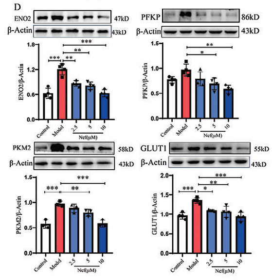
Disruption of the eEF1A1/ARID3A/PKC-δ Complex by Neferine Inhibits Macrophage Glycolytic Reprogramming in Atherosclerosis
Author: Baoping Xie, Li-Wen Tian, Chenxu Liu, Jiahua Li, Xiaoyu Tian, Rong Zhang, Fan Zhang, Zhongqiu Liu, Yuanyuan Cheng
PMID: 39973763

Journal: Advanced Science
アプリケーション: WB
交差性: Mouse
掲載日: 2025 Feb
-
Citation
-
From COPD to cancer: indacaterol’s unexpected role in combating NSCLC
Author: Chenghao Liu, Jiaqi Huang, Pengjie Cai, Min Jiang, Honglei Chen
PMID: 40276602
Journal: Frontiers In Pharmacology
アプリケーション: IHC-P,IF
交差性: Mouse,Human
掲載日: 2025 Apr
-
Citation
-
Ochratoxin A-induced mitochondrial pathway apoptosis and ferroptosis by promoting glycolysis
Author: Zhou Yao, Chen Wenying, Feng Shiyu, Liu Shuangchao, Chen Cheng, Yao Bingxu, Shen Xiao Li
PMID: 40167953
Journal: Apoptosis
アプリケーション: WB
交差性: Human
掲載日: 2025 Apr
-
Citation
-
Metabolic Reprogramming of CD4+ T Cells by Mesenchymal Stem Cell-Derived Extracellular Vesicles Attenuates Autoimmune Hepatitis Through Mitochondrial Protein Transfer
Author: Mengyi Shen, Leyu Zhou, Xiaoli Fan, Ruiqi Wu, Shuyun Liu, Qiaoyu Deng, Yanyi Zheng, Jingping Liu, Li Yang
PMID: 39345912
Journal: International Journal Of Nanomedicine
アプリケーション: WB
交差性: Mouse
掲載日: 2024 Sept
-
Citation
-
Shikonin protects mitochondria through the NFAT5/AMPK pathway for the treatment of diabetic wounds
Author: Lu-Sha Cen,et al
PMID: 39676806
Journal: World Journal Of Diabetes
アプリケーション: WB
交差性: Human
掲載日: 2024 Nov
-
Citation
-
Astrocytic LRP1 enables mitochondria transfer to neurons and mitigates brain ischemic stroke by suppressing ARF1 lactylation
Author: Zhou Jian,et al
PMID: 38906140

Journal: Cell Metabolism
アプリケーション: IF
交差性: Mouse
掲載日: 2024 Jun
-
Citation
-
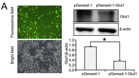
“Golgi-customized Trojan horse” nanodiamonds impair GLUT1 plasma membrane localization and inhibit tumor glycolysis
Author: Bei Kang, Haobo Wang, Huaqing Jing, Yunsheng Dou, Sona Krizkova, Zbynek Heger, Vojtech Adam, Nan Li
PMID: 38789089
Journal: Journal Of Controlled Release
アプリケーション:
交差性:
掲載日: 2024 Jun
-
Citation
-
Low-level laser therapy alleviates periodontal age-related inflammation in diabetic mice via the GLUT1/mTOR pathway
Author: Cui Aimin,et al
PMID: 38236306
Journal: Journal Of Lasers In Medical Sciences
アプリケーション: IF
交差性: Mouse
掲載日: 2024 Jan
-
Citation
-
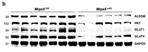
A defect in mitochondrial protein translation influences mitonuclear communication in the heart
Author: Gao, F., Liang, T., Lu, Y. W., Fu, X., Dong, X., Pu, L., Hong, T., Zhou, Y., Zhang, Y., Liu, N., Zhang, F., Liu, J., Malizia, A. P., Yu, H., Zhu, W., Cowan, D. B., Chen, H., Hu, X., Mably, J. D., Wang, J., … Chen, J.
PMID: 36949106

Journal: Nature Communications
アプリケーション: WB
交差性: Mouse
掲載日: 2023 Mar
-
Citation
-
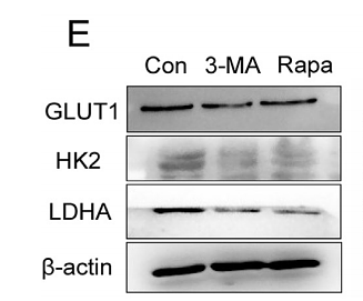
Autophagy participates in germline cyst breakdown and follicular formation by modulating glycolysis switch via Akt signaling in newly-hatched chicken ovaries
Author:
PMID: 35525303

Journal: Developmental Biology
アプリケーション: IF-tissue,WB
交差性: Chicken
掲載日: 2022 Jul
-
Citation
-
Metabolic heterogeneity caused by HLA-DRB1*04:05 and protective effect of inosine on autoimmune hepatitis
Author: Yang, F., Zhou, L., Shen, Y., Zhao, S., Zheng, Y., Men, R., Fan, X., & Yang, L.
PMID: 35990653

Journal: Frontiers In Immunology
アプリケーション: WB
交差性: Mouse
掲載日: 2022 Aug
-
Citation
-
DNA demethylase ALKBH1 promotes adipogenic differentiation via regulation of HIF-1 signaling
Author: Liu, Y., Chen, Y., Wang, Y., Jiang, S., Lin, W., Wu, Y., Li, Q., Guo, Y., Liu, W., & Yuan, Q.
PMID: 34922943

Journal: Journal Of Biological Chemistry
アプリケーション: WB
交差性: Human
掲載日: 2021 Jan
-
Citation
-
Activation of AMPK pathway compromises Rab11 downregulation-mediated inhibition of Schwann cell proliferation in a Glut1 and Glut3-dependent manner
Author:
PMID: 31954765

Journal: Neuroscience Letters
アプリケーション: WB,IF-Cell
交差性: Rat
掲載日: 2020 Feb
-
Citation
同じターゲット & 同じ経路の製品
Glucose Transporter GLUT1 Recombinant Antibody [SA0377] - Rat IgG1 (Chimeric) - BSA and Azide free
Application: IHC-Fr,IHC-P,IF-Tissue,WB
Reactivity: Human,Mouse,Rat
Conjugate: unconjugated
Glucose Transporter GLUT1 Recombinant Antibody [SA0377] - Rat IgG1 (Chimeric)
Application: IHC-Fr,IHC-P,IF-Tissue,WB
Reactivity: Human,Mouse,Rat
Conjugate: unconjugated
Glucose Transporter GLUT1 Recombinant Rabbit Monoclonal Antibody [SA0377] - BSA and Azide free
Application: WB,IF-Cell,IF-Tissue,IHC-P,FC
Reactivity: Human,Mouse,Rat
Conjugate: unconjugated
Glucose Transporter GLUT1 Recombinant Rabbit Monoclonal Antibody
Application: mIHC
Reactivity: Human
Conjugate: unconjugated
Glucose Transporter GLUT1 Rabbit Polyclonal Antibody
Application: WB,IF-Cell,IHC-P,FC
Reactivity: Human,Mouse
Conjugate: unconjugated
iFluor™ 488 Conjugated Glucose Transporter GLUT1 Recombinant Rabbit Monoclonal Antibody [SA0377]
Application: IF-Cell,IF-Tissue
Reactivity: Human,Mouse,Rat
Conjugate: iFluor™ 488
iFluor™ 647 Conjugated Glucose Transporter GLUT1 Recombinant Rabbit Monoclonal Antibody [SA0377]
Application: IF-Tissue
Reactivity: Human,Mouse
Conjugate: iFluor™ 647


