Vinculin Recombinant Rabbit Monoclonal Antibody [JM42-43]

製品仕様
今すぐ注文,【现货】
問い合わせをクリックCatalog# ET1705-94
Vinculin Recombinant Rabbit Monoclonal Antibody [JM42-43]
-
WB
-
IF-Cell
-
IF-Tissue
-
IHC-P
-
Human
-
Mouse
-
Rat
-
HA750448
不含抗保成分
-
ET1705-94
含抗保成分
-
unconjugated
概要
製品名
Vinculin Recombinant Rabbit Monoclonal Antibody [JM42-43]
抗体の種類
Recombinant Rabbit monoclonal Antibody
免疫原
Synthetic peptide within human Vinculin aa 1,000-1,060.
種属反応性
Human, Mouse, Rat
検証された応用例
WB, IF-Cell, IF-Tissue, IHC-P
分子量
Predicted band size: 124 kDa
陽性対照
HepG2 cell lysate, HeLa cell lysate, NIH/3T3 cell lysate, C6 cell lysate, Mouse spleen tissue lysate, Rat kidney tissue lysate, NIH/3T3, C6, HeLa, mouse spleen tissue, mouse testis tissue, rat colon tissue, rat spleen tissue, rat testis tissue.
結合
unconjugated
クローン番号
JM42-43
RRID
製品の特徴
形態
Liquid
保存方法
Shipped at 4℃. Store at +4℃ short term (1-2 weeks). It is recommended to aliquot into single-use upon delivery. Store at -20℃ long term.
保存用バッファー
1*TBS (pH7.4), 0.05% BSA, 40% Glycerol. Preservative: 0.05% Sodium Azide.
アイソタイプ
IgG
精製方法
Protein A affinity purified.
応用希釈度
-
WB
-
1:20,000-1:50,000
-
IF-Cell
-
1:200-1:1,000
-
IF-Tissue
-
1:500-1:4,000
-
IHC-P
-
1:5,000-1:20,000
論文における応用例
| WB | 確認する 14 以下の論文 |
| IF | 確認する 3 以下の論文 |
| IF-Cell | 確認する 2 以下の論文 |
| IHC-P | 確認する 1 以下の論文 |
論文における種属
| Human | 確認する 11 以下の論文 |
| Mouse | 確認する 10 以下の論文 |
| Rat | 確認する 2 以下の論文 |
| Zebrafish | 確認する 1 以下の論文 |
ターゲット
機能
Focal adhesions are identified as areas within the plasma membrane of tissue culture cells that adhere tightly to the underlying substrate. In vivo, these regions are involved in the adhesion of cells to the extracellular matrix. Paxillin and vinculin are cytoskeletal, focal adhesion proteins that are components of a protein complex which links the Actin network to the plasma membrane. Vinculin binding sites have been identified on other cytoskeletal proteins, including Talin and α-actinin. In addition, vinculin, Talin and α-actinin each contain Actin binding sites. Expression of vinculin and Talin have been shown to be affected by the level of Actin expression. α-Actinin has been shown to link Actin to integrins in the plasma membrane through interactions with the vinculin and Talin complex or by a direct interaction with integrin.
背景文献
1. King HO et al. RAD51 Is a Selective DNA Repair Target to Radiosensitize Glioma Stem Cells. Stem Cell Reports 8:125-139 (2017).
2. Boyette LB et al. Phenotype, function, and differentiation potential of human monocyte subsets. PLoS One 12:e0176460 (2017).
配列相同性
Belongs to the vinculin/alpha-catenin family.
組織特異性
Metavinculin is muscle-specific.
翻訳後修飾
Phosphorylated; on serines, threonines and tyrosines. Phosphorylation on Tyr-1133 in activated platelets affects head-tail interactions and cell spreading but has no effect on actin binding nor on localization to focal adhesion plaques (By similarity).; Acetylated; mainly by myristic acid but also by a small amount of palmitic acid.
亜細胞局在
Cell junction, Cell membrane, Cytoplasm, Cytoskeleton, Membrane.
別名
CMD1W antibody
CMH15 antibody
Epididymis luminal protein 114 antibody
HEL114 antibody
Metavinculin antibody
MV antibody
MVCL antibody
OTTHUMP00000019861 antibody
OTTHUMP00000019862 antibody
VCL antibody
展開CMD1W antibody
CMH15 antibody
Epididymis luminal protein 114 antibody
HEL114 antibody
Metavinculin antibody
MV antibody
MVCL antibody
OTTHUMP00000019861 antibody
OTTHUMP00000019862 antibody
VCL antibody
VINC antibody
VINC_HUMAN antibody
Vinculin antibody
折りたたむ画像
-

Western blot analysis of Vinculin on different lysates with Rabbit anti-Vinculin antibody (ET1705-94) at 1/20,000 dilution.
Lane 1: HepG2 cell lysate (15 µg/Lane)
Lane 2: HeLa cell lysate (15 µg/Lane)
Lane 3: NIH/3T3 cell lysate (15 µg/Lane)
Lane 4: C6 cell lysate (15 µg/Lane)
Lane 5: Mouse spleen tissue lysate (20 µg/Lane)
Lane 6: Rat kidney tissue lysate (20 µg/Lane)
Predicted band size: 124 kDa
Observed band size: 124 kDa
Exposure time: 1 minute 21 seconds;
4-20% SDS-PAGE gel.
Proteins were transferred to a PVDF membrane and blocked with 5% NFDM/TBST for 1 hour at room temperature. The primary antibody (ET1705-94) at 1/20,000 dilution was used in 5% NFDM/TBST at 4℃ overnight. Goat Anti-Rabbit IgG - HRP Secondary Antibody (HA1001) at 1/50,000 dilution was used for 1 hour at room temperature. -

☑ Knockdown (KD)
Western blot analysis of Vinculin on different lysates with Rabbit anti-Vinculin antibody (ET1705-94) at 1/20,000 dilution.
Lane 1: HAP1-parental cell lysate
Lane 2: HAP1-Vinculin KD cell lysate
Lysates/proteins at 10 µg/Lane.
Predicted band size: 124 kDa
Observed band size: 124 kDa
Exposure time: 10 seconds; ECL: K1801;
4-20% SDS-PAGE gel.
Proteins were transferred to a PVDF membrane and blocked with 5% NFDM/TBST for 1 hour at room temperature. The primary antibody (ET1705-94) at 1/20,000 dilution was used in K1803 at 4℃ overnight. Goat Anti-Rabbit IgG - HRP Secondary Antibody (HA1001) at 1/50,000 dilution was used for 1 hour at room temperature. -

Immunocytochemistry analysis of NIH/3T3 cells labeling Vinculin with Rabbit anti-Vinculin antibody (ET1705-94) at 1/250 dilution.
Cells were fixed in 4% paraformaldehyde for 20 minutes at room temperature, permeabilized with 0.1% Triton X-100 in PBS for 5 minutes at room temperature, then blocked with 1% BSA in 10% negative goat serum for 1 hour at room temperature. Cells were then incubated with Rabbit anti-Vinculin antibody (ET1705-94) at 1/1250 dilution in 1% BSA in PBST overnight at 4 ℃. Goat Anti-Rabbit IgG H&L (iFluor™ 488, HA1121) was used as the secondary antibody at 1/1,000 dilution. PBS instead of the primary antibody was used as the secondary antibody only control. Nuclear DNA was labelled in blue with DAPI.
Beta tubulin (M1305-2, red) was stained at 1/100 dilution overnight at +4℃. Goat Anti-Mouse IgG H&L (iFluor™ 594, HA1126) was used as the secondary antibody at 1/1,000 dilution. -

Immunocytochemistry analysis of C6 cells labeling Vinculin with Rabbit anti-Vinculin antibody (ET1705-94) at 1/250 dilution.
Cells were fixed in 4% paraformaldehyde for 20 minutes at room temperature, permeabilized with 0.1% Triton X-100 in PBS for 5 minutes at room temperature, then blocked with 1% BSA in 10% negative goat serum for 1 hour at room temperature. Cells were then incubated with Rabbit anti-Vinculin antibody (ET1705-94) at 1/1250 dilution in 1% BSA in PBST overnight at 4 ℃. Goat Anti-Rabbit IgG H&L (iFluor™ 488, HA1121) was used as the secondary antibody at 1/1,000 dilution. PBS instead of the primary antibody was used as the secondary antibody only control. Nuclear DNA was labelled in blue with DAPI.
Beta tubulin (M1305-2, red) was stained at 1/100 dilution overnight at +4℃. Goat Anti-Mouse IgG H&L (iFluor™ 594, HA1126) was used as the secondary antibody at 1/1,000 dilution. -

Immunocytochemistry analysis of HeLa cells labeling Vinculin with Rabbit anti-Vinculin antibody (ET1705-94) at 1/1,000 dilution.
Cells were fixed in 100% precooled methanol for 5 minutes at room temperature, then blocked with 1% BSA in 10% negative goat serum for 1 hour at room temperature. Cells were then incubated with Rabbit anti-Vinculin antibody (ET1705-94) at 1/1,000 dilution in 1% BSA in PBST overnight at 4 ℃. Goat Anti-Rabbit IgG H&L (iFluor™ 488, HA1121) was used as the secondary antibody at 1/1,000 dilution. PBS instead of the primary antibody was used as the secondary antibody only control. Nuclear DNA was labelled in blue with DAPI. -

Immunohistochemical analysis of paraffin-embedded mouse spleen tissue with Rabbit anti-Vinculin antibody (ET1705-94) at 1/20,000 dilution.
The section was pre-treated using heat mediated antigen retrieval with Tris-EDTA buffer (pH 9.0) for 20 minutes. The tissues were blocked in 1% BSA for 20 minutes at room temperature, washed with ddH2O and PBS, and then probed with the primary antibody (ET1705-94) at 1/20,000 dilution for 1 hour at room temperature. The detection was performed using an HRP conjugated compact polymer system. DAB was used as the chromogen. Tissues were counterstained with hematoxylin and mounted with DPX. -

Immunohistochemical analysis of paraffin-embedded mouse testis tissue with Rabbit anti-Vinculin antibody (ET1705-94) at 1/20,000 dilution.
The section was pre-treated using heat mediated antigen retrieval with Tris-EDTA buffer (pH 9.0) for 20 minutes. The tissues were blocked in 1% BSA for 20 minutes at room temperature, washed with ddH2O and PBS, and then probed with the primary antibody (ET1705-94) at 1/20,000 dilution for 1 hour at room temperature. The detection was performed using an HRP conjugated compact polymer system. DAB was used as the chromogen. Tissues were counterstained with hematoxylin and mounted with DPX. -

Immunohistochemical analysis of paraffin-embedded rat colon tissue with Rabbit anti-Vinculin antibody (ET1705-94) at 1/20,000 dilution.
The section was pre-treated using heat mediated antigen retrieval with Tris-EDTA buffer (pH 9.0) for 20 minutes. The tissues were blocked in 1% BSA for 20 minutes at room temperature, washed with ddH2O and PBS, and then probed with the primary antibody (ET1705-94) at 1/20,000 dilution for 1 hour at room temperature. The detection was performed using an HRP conjugated compact polymer system. DAB was used as the chromogen. Tissues were counterstained with hematoxylin and mounted with DPX. -

Immunohistochemical analysis of paraffin-embedded rat spleen tissue with Rabbit anti-Vinculin antibody (ET1705-94) at 1/20,000 dilution.
The section was pre-treated using heat mediated antigen retrieval with Tris-EDTA buffer (pH 9.0) for 20 minutes. The tissues were blocked in 1% BSA for 20 minutes at room temperature, washed with ddH2O and PBS, and then probed with the primary antibody (ET1705-94) at 1/20,000 dilution for 1 hour at room temperature. The detection was performed using an HRP conjugated compact polymer system. DAB was used as the chromogen. Tissues were counterstained with hematoxylin and mounted with DPX. -

Immunohistochemical analysis of paraffin-embedded rat testis tissue with Rabbit anti-Vinculin antibody (ET1705-94) at 1/20,000 dilution.
The section was pre-treated using heat mediated antigen retrieval with Tris-EDTA buffer (pH 9.0) for 20 minutes. The tissues were blocked in 1% BSA for 20 minutes at room temperature, washed with ddH2O and PBS, and then probed with the primary antibody (ET1705-94) at 1/20,000 dilution for 1 hour at room temperature. The detection was performed using an HRP conjugated compact polymer system. DAB was used as the chromogen. Tissues were counterstained with hematoxylin and mounted with DPX. -

Flow cytometric analysis of HeLa cells labeling Vinculin.
Cells were fixed and permeabilized. Then stained with the primary antibody (ET1705-94, 1μg/mL) (red) compared with Rabbit IgG Isotype Control (green). After incubation of the primary antibody at +4℃ for an hour, the cells were stained with a iFluor™ 488 conjugate-Goat anti-Rabbit IgG Secondary antibody (HA1121) at 1/1,000 dilution for 30 minutes at +4℃. Unlabelled sample was used as a control (cells without incubation with primary antibody; black).
ご注意ください: All products are "FOR RESEARCH USE ONLY AND ARE NOT INTENDED FOR DIAGNOSTIC OR THERAPEUTIC USE"
論文での実績
-
Metformin alleviates ribociclib-induced lung injury by restoring impaired autophagy via targeting Mucolipin-1
Author: Yueping Qiu, Jing Chen, Yu Zhang, Chang Shu, Yan Hu, Libin Pan, Wenxiu Xin, Haiying Ding, Shuanghui Lu, Luo Fang
PMID: 10.1016/j.taap.2025.117630
Journal: Toxicology And Applied Pharmacology
アプリケーション: WB
交差性: Human
掲載日: 2025 Nov
-
Citation
-
Sappanchalcone Suppresses NSCLC by Oxidative Stress-Driven DNA Damage and ER Stress Activation through PIEZO1 Modulation
Author: Weiyu Wu, Ren Zhang, Geer Chen, Ziyu Chen, Zicong Lin, Yin Chen, Jiaqi Li, Weilin Liao, Junyi Wang, Xiaoxuan Wang, Junhao Huang, Lijuan Ma, Haijie Yu
PMID: 10.1016/j.isci.2025.114057
Journal: iScience
アプリケーション: WB
交差性: Human
掲載日: 2025 Nov
-
Citation
-
Mussel-inspired bifunctional chimeric peptides macromolecules functionalize 3D-printed porous scaffolds for enhanced antimicrobial and osseointegration properties in bone defect repair
Author: Ziyang Bai, Yifan Zhao, Wenjun Zhang, Chenying Cui, Jingyu Yan, Meijun Du, Jiahui Tong, Yingyu Liu, Ying Zhang, Ke Zhang, Binbin Zhang, Xia Li, Xiuping Wu, Bing Li
PMID: 40174844
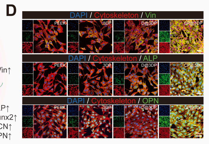
Journal: International Journal Of Biological Macromolecules
アプリケーション: IF
交差性: Rat
掲載日: 2025 Mar
-
Citation
-
ROS-Responsive Hydrogel for Localized Delivery of Nampt and Stat3 Inhibitors Exhibits Synergistic Antitumor Effects in Colorectal Cancer Through Ferroptosis Induction and Immune Microenvironment Remodeling
Author: Chenyang Ye, Mi Mi, Saimeng Shi, Lina Qi, Shanshan Weng, Lu Wang, Yier Lu, Chao Chen, Yinuo Tan, Mengyuan Yang, Cheng Guo, Rui Bai, Xuefeng Fang, Ji Wang, Ying Yuan
PMID: 40492508
Journal: Advanced Science
アプリケーション: WB
交差性: Mouse
掲載日: 2025 Jun
-
Citation
-
Targeting MAN1B1 potently enhances bladder cancer antitumor immunity via deglycosylation of CD47
Author: Jie Zhang, Chen Zhang, Ruichen Zang, Weiwu Chen, Yining Guo, Haofei Jiang, Jing Le, Kunyu Wang, Haobo Fan, Xudong Wang, Sisi Mo, Peng Gao, Wenhao Guo, Xinrong Jiang, Fengbin Gao, Junming Jiang, Juyan Zheng, Yuxing Chen, Yicheng Chen, Yanlan Yu, Guoqing Ding
PMID: 40493414
Journal: Cancer Communications
アプリケーション: WB
交差性: Human,Mouse
掲載日: 2025 Jun
-
Citation
-
A Novel Missense Variant of BMPR1A in Juvenile Polyposis Syndrome: Assessment of Structural and Functional Alternations
Author: Mengyuan Yang, Ziyan Tong, Zhijun Yuan, Bingjing Jiang, Yingxin Zhao, Dong Xu, Ying Yuan
PMID: 40226309
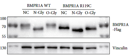
Journal: Human Mutation
アプリケーション: WB
交差性: Human
掲載日: 2025 Feb
-
Citation
-
The TIM22 carrier translocase supports cell proliferation by facilitating mitochondrial iron uptake for Fe-S biogenesis
Author: Shuai Liu, Qingyu Li, Mengye Cao, Zefang Bimo Zhao, Cong Liu, Zhuoran Zhen, Jiankun Ren, Chengyang Liu, Danyang Ruan, Luna Zhang, Wenjuan Zhang, Hao Gong, Xiaolong Liu, Xuejie Zhang, Deng Pan, Weijun Pan, Jiajun Zhu
PMID: 10.1016/j.molcel.2025.11.022
Journal: Molecular Cell
アプリケーション: WB
交差性: Zebrafish,Human
掲載日: 2025 Dec
-
Citation
-
Glabridin-Gold(I) Complex as a Novel Immunomodulatory Agent Targeting TrxR and MAPK Pathways for Synergistic Enhancement of Antitumor Immunity
Author: Zhaoran Wang, Meiyu Wang, Qiong Chen, Mengshi Wang, Fuwei Li, Lin Lv, Zhenfan Wen, Zhongren Xu, Yixia Yang, Chunyang Bi, Wukun Liu
PMID: 40842087
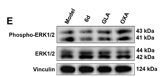
Journal: Advanced Science
アプリケーション: WB
交差性: Mouse,Human
掲載日: 2025 Aug
-
Citation
-
MXene-inducing self-assembled Bombyx mori (B. mori) silk fibroin nanofibers improve the early cell adhesion and neuronal differentiation of neural stem cells
Author: Zhangze Yang, Meng Zhang, Yi Wu, Jiaping Xu, Jie Wang, Quan Wan, Zongpu Xu, Yajun Shuai, Junhui Lv, Jiaqi Hu, Chuanbin Mao, Shuxu Yang, Mingying Yang
PMID: 12NOPMID25092501
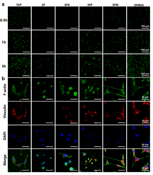
Journal: Chemical Engineering Journal
アプリケーション: IF
交差性: Rat
掲載日: 2025 Aug
-
Citation
-
LAMA4+ CD90+ eCAFs provide immunosuppressive microenvironment for liver cancer through induction of CD8+ T cell senescence
Author: Zhang Jianlei, Li Zhihui, Zhang Qiong, Ma Wen, Fan Weina, Dong Jing, Tian Jingjie, Liao Hongfan, Guo Junzhe, Cao Yabing, Yin Jiang, Zheng Guopei, Li Nan
PMID: 40289085
Journal: Cell Communication And Signaling
アプリケーション: WB
交差性: Mouse,Human
掲載日: 2025 Apr
-
Citation
-
Developing patient-derived organoids to identify JX24120 inhibit SAMe synthesis in endometrial cancer by targeting MAT2B
Author: Chunxue Zhang,et al
PMID: 39293586
Journal: Pharmacological Research
アプリケーション: WB
交差性: Mouse
掲載日: 2024 Sep
-
Citation
-
Nicotine promotes Staphylococcus aureus-induced osteomyelitis by activating the Nrf2/Slc7a11 signaling axis
Author: Zhou Xuyou,et al
PMID: 38772295
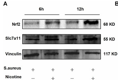
Journal: International Immunopharmacology
アプリケーション: WB
交差性: Mouse
掲載日: 2024 May
-
Citation
-
DIREN mitigates DSS-induced colitis in mice and attenuates collagen deposition via inhibiting the Wnt/β-catenin and focal adhesion pathways
Author: Lai Weizhi,et al
PMID: 38678963
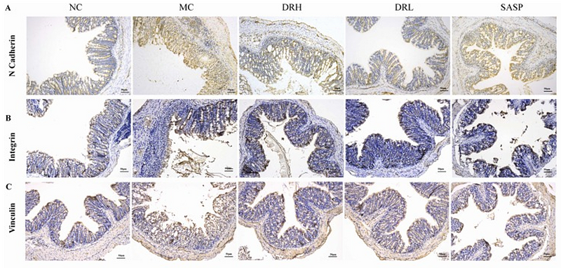
Journal: Biomedicine & Pharmacotherapy
アプリケーション: IHC-P
交差性: Mouse
掲載日: 2024 Apr
-
Citation
-
MC3T3-E1 cells lead to bone loss in Staphylococcus aureus osteomyelitis through oxeiptosis pathway
Author: Xu Yuan,et al
PMID: no pmid0514
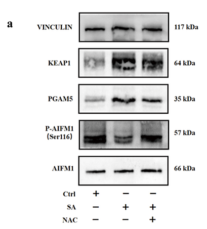
Journal: Preprint And Has Not Been Certified By Peer Review
アプリケーション: WB
交差性: Mouse
掲載日: 2024 Apr
-
Citation
-
TWIST1 rescue calcium overload and apoptosis induced by inflammatory microenvironment in S. aureus-induced osteomyelitis
Author:
PMID: 37071966
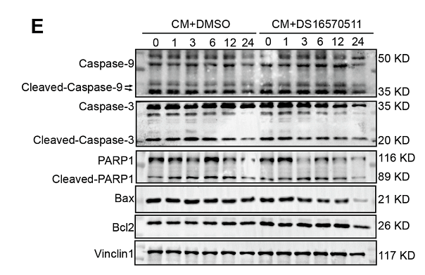
Journal: International Immunopharmacology
アプリケーション: WB
交差性: Mouse
掲載日: 2023 Jun
-
Citation
-
Enhancing cell adhesive and antibacterial activities of glass-fibre-reinforced polyetherketoneketone through Mg and Ag PIII
Author:
PMID: 37489146
Journal: Regenerative Biomaterials
アプリケーション: IF
交差性:
掲載日: 2023 Jul
-
Citation
-
Effects of matrix viscoelasticity on cell–matrix interaction, actin cytoskeleton organization, and apoptosis of osteosarcoma MG-63 cells
Author: Deng Huan,et al
PMID: 38079114
Journal: Journal Of Materials Chemistry B
アプリケーション: IF-Cell
交差性: Human
掲載日: 2023 Dec
-
Citation
-
Matrix Stiffness Regulated Endoplasmic Reticulum Stress-mediated Apoptosis of Osteosarcoma Cell through Ras Signal Cascades
Author: Deng Huan,et al
PMID: 37789235
Journal: Cell Biochemistry And Biophysics
アプリケーション: IF-Cell
交差性: Human,Mouse
掲載日: 2023 Dec
-
Citation
-
Targeting eIF3f Suppresses the Growth of Prostate Cancer Cells by Inhibiting Akt Signaling
Author: Guowei Shi
PMID: 32440143
Journal: Onco Targets And Therapy
アプリケーション: WB
交差性: Human
掲載日: 2020 May
-
Citation
-
Ang II-AT2R increases mesenchymal stem cell migration by signaling through the FAK and RhoA/Cdc42 pathways in vitro
Author: Hai-bo Qiu
PMID: 28697804
Journal: Stem Cell Research & Therapy
アプリケーション: WB
交差性: Human
掲載日: 2017 Jul
-
Citation
同じターゲット & 同じ経路の製品
Vinculin Recombinant Rabbit Monoclonal Antibody [JM42-43] - BSA and Azide free
Application: WB,IF-Cell,IF-Tissue,IHC-P
Reactivity: Human,Mouse,Rat
Conjugate: unconjugated


