TSG101 Recombinant Rabbit Monoclonal Antibody [JJ0900]

製品仕様
今すぐ注文,【现货】
問い合わせをクリックCatalog# ET1701-59
TSG101 Recombinant Rabbit Monoclonal Antibody [JJ0900]
-
WB
-
IF-Cell
-
IF-Tissue
-
IHC-P
-
FC
-
Human
-
Mouse
-
Rat
概要
製品名
TSG101 Recombinant Rabbit Monoclonal Antibody [JJ0900]
抗体の種類
Recombinant Rabbit monoclonal Antibody
免疫原
Synthetic peptide within Human TSG101 aa 24-73 / 390.
種属反応性
Human, Mouse, Rat
検証された応用例
WB, IF-Cell, IF-Tissue, IHC-P, FC
分子量
Predicted band size: 44 kDa
陽性対照
HeLa cell lysate, K-562 cell lysate, MCF7 cell lysate, Jurkat cell lysate, C2C12 cell lysate, PC-12 cell lysate, NIH/3T3 cell lysate, mouse brain tissue lysate, rat brain tissue lysate, HeLa, NIH/3T3, PC-12, human kidney tissue, mouse colon tissue, mouse kidney tissue, human colon carcinoma tissue.
結合
unconjugated
クローン番号
JJ0900
RRID
製品の特徴
形態
Liquid
保存方法
Shipped at 4℃. Store at +4℃ short term (1-2 weeks). It is recommended to aliquot into single-use upon delivery. Store at -20℃ long term.
保存用バッファー
1*TBS (pH7.4), 0.05% BSA, 40% Glycerol. Preservative: 0.05% Sodium Azide.
アイソタイプ
IgG
精製方法
Protein A affinity purified.
応用希釈度
-
WB
-
1:2,000-1:5,000
-
IF-Cell
-
1:200
-
IF-Tissue
-
1:200-1:500
-
IHC-P
-
1:500-1:1,000
-
FC
-
1:1,000
論文における応用例
| WB | 確認する 37 以下の論文 |
| IF | 確認する 3 以下の論文 |
| IHC-P | 確認する 2 以下の論文 |
| IF-cell | 確認する 1 以下の論文 |
論文における種属
| Human | 確認する 18 以下の論文 |
| Mouse | 確認する 15 以下の論文 |
| Rat | 確認する 4 以下の論文 |
| Turtle | 確認する 1 以下の論文 |
| zebrafish | 確認する 1 以下の論文 |
| Rabbit | 確認する 1 以下の論文 |
| Goat | 確認する 1 以下の論文 |
ターゲット
機能
The transformation of a normal cell to one that is malignant can result from mutations in genes that encode proteins with key regulatory functions. Examples include the retinoblastoma gene product (Rb p110), p53, VHL and APC. Using a novel cloning strategy that allows the isolation of previously uncharacterized genes encoding selectable recessive phenotypes, an additional tumor suppressor gene has been identified. This gene, termed tsg 101 for tumor susceptibility gene 101, encodes a stathmin binding domain protein. When expression of this growth inhibitory gene is blocked in NIH/3T3 cells using antisense mRNA, the cells exhibit a transformed phenotype and are tumorigenic in SL6 mice.
背景文献
1. Ruiz-Guillen M et al. Capsid-deficient alphaviruses generate propagative infectious microvesicles at the plasma membrane. Cell Mol Life Sci 73:3897-916 (2016).
2. Baranyai T et al. Isolation of Exosomes from Blood Plasma: Qualitative and Quantitative Comparison of Ultracentrifugation and Size Exclusion Chromatography Methods. PLoS One 10:e0145686 (2015).
配列相同性
Belongs to the ubiquitin-conjugating enzyme family. UEV subfamily.
組織特異性
Heart, brain, placenta, lung, liver, skeletal, kidney and pancreas.
翻訳後修飾
Monoubiquitinated at multiple sites by LRSAM1 and by MGRN1. Ubiquitination inactivates it, possibly by regulating its shuttling between an active membrane-bound protein and an inactive soluble form. Ubiquitination by MGRN1 requires the presence of UBE2D1.
亜細胞局在
Cytoplasm, Cytoskeleton, Endosome, Membrane, Nucleus.
別名
ESCRT I complex subunit TSG101 antibody
ESCRT-I complex subunit TSG101 antibody
TS101_HUMAN antibody
TSG 10 antibody
TSG 101 antibody
TSG10 antibody
Tsg101 antibody
Tumor susceptibility gene 10 antibody
Tumor susceptibility gene 101 antibody
Tumor susceptibility gene 101 protein antibody
展開ESCRT I complex subunit TSG101 antibody
ESCRT-I complex subunit TSG101 antibody
TS101_HUMAN antibody
TSG 10 antibody
TSG 101 antibody
TSG10 antibody
Tsg101 antibody
Tumor susceptibility gene 10 antibody
Tumor susceptibility gene 101 antibody
Tumor susceptibility gene 101 protein antibody
Tumor susceptibility protein antibody
Tumor susceptibility protein isoform 3 antibody
VPS 23 antibody
VPS23 antibody
折りたたむ画像
-

Western blot analysis of TSG101 on different lysates with Rabbit anti-TSG101 antibody (ET1701-59) at 1/2,000 dilution and competitor's antibody at 1/1,000 dilution.
Lane 1: HeLa cell lysate
Lane 2: K-562 cell lysate
Lane 3: MCF7 cell lysate
Lane 4: Jurkat cell lysate
Lane 5: C2C12 cell lysate
Lane 6: PC-12 cell lysate
Lysates/proteins at 20 µg/Lane.
Predicted band size: 44 kDa
Observed band size: 44/47 kDa
Exposure time: 3 minutes 20 seconds; ECL: K1801;
4-20% SDS-PAGE gel.
Proteins were transferred to a PVDF membrane and blocked with 5% NFDM/TBST for 1 hour at room temperature. The primary antibody (ET1701-59) at 1/2,000 dilution and competitor's antibody at 1/1,000 dilution were used in 5% NFDM/TBST at 4℃ overnight. Goat Anti-Rabbit IgG - HRP Secondary Antibody (HA1001) at 1/50,000 dilution was used for 1 hour at room temperature. -
![<span style="font-weight: bold;">☑ Knockdown (KD)</span><br /><br />Western blot analysis of TSG101 with anti-TSG101 antibody[JJ0900] (<a href="/products/ET1701-59" style="font-weight: bold;text-decoration: underline;">ET1701-59</a>) at 1/2,000 dilution.<br />Lane 1: Wild-type Hela whole cell lysate (10 µg).<br />Lane 2/3: TSG101 knockdown Hela whole cell lysate (10 µg).<br /><br />Proteins were transferred to a PVDF membrane and blocked with 5% NFDM in TBST for 1 hour at room temperature. The primary antibody (<a href="/products/ET1701-59" style="font-weight: bold;text-decoration: underline;">ET1701-59</a>, 1/2,000) was used in 5% BSA at room temperature for 2 hours. Goat Anti-Rabbit IgG-HRP Secondary Antibody (<a href="/products/HA1001" style="font-weight: bold;text-decoration: underline;">HA1001</a>) at 1/50,000 dilution was used for 1 hour at room temperature.](https://storage.huabio.cn/huabio/productImg/ET1701-59_2.jpg?v=20250328162426)
☑ Knockdown (KD)
Western blot analysis of TSG101 with anti-TSG101 antibody[JJ0900] (ET1701-59) at 1/2,000 dilution.
Lane 1: Wild-type Hela whole cell lysate (10 µg).
Lane 2/3: TSG101 knockdown Hela whole cell lysate (10 µg).
Proteins were transferred to a PVDF membrane and blocked with 5% NFDM in TBST for 1 hour at room temperature. The primary antibody (ET1701-59, 1/2,000) was used in 5% BSA at room temperature for 2 hours. Goat Anti-Rabbit IgG-HRP Secondary Antibody (HA1001) at 1/50,000 dilution was used for 1 hour at room temperature. -

Western blot analysis of TSG101 on different lysates with Rabbit anti-TSG101 antibody (ET1701-59) at 1/2,000 dilution.
Lane 1: HeLa cell lysate
Lane 2: K-562 cell lysate
Lane 3: Jurkat cell lysate
Lane 4: NIH/3T3 cell lysate
Lane 5: PC-12 cell lysate
Lane 6: Mouse brain tissue lysate
Lane 7: Rat brain tissue lysate
Lysates/proteins at 20 µg/Lane.
Predicted band size: 44 kDa
Observed band size: 44/47 kDa
Exposure time: 1 minute 40 seconds; ECL: K1801;
4-20% SDS-PAGE gel.
Proteins were transferred to a PVDF membrane and blocked with 5% NFDM/TBST for 1 hour at room temperature. The primary antibody (ET1701-59) at 1/2,000 dilution was used in 5% NFDM/TBST at 4℃ overnight. Goat Anti-Rabbit IgG - HRP Secondary Antibody (HA1001) at 1/50,000 dilution was used for 1 hour at room temperature. -

Immunocytochemistry analysis of HeLa cells labeling TSG101 with Rabbit anti-TSG101 antibody (ET1701-59) at 1/200 dilution.
Cells were fixed in 4% paraformaldehyde for 20 minutes at room temperature, permeabilized with 0.1% Triton X-100 in PBS for 5 minutes at room temperature, then blocked with 1% BSA in 10% negative goat serum for 1 hour at room temperature. Cells were then incubated with Rabbit anti-TSG101 antibody (ET1701-59) at 1/200 dilution in 1% BSA in PBST overnight at 4 ℃. Goat Anti-Rabbit IgG H&L (iFluor™ 488, HA1121) was used as the secondary antibody at 1/1,000 dilution. PBS instead of the primary antibody was used as the secondary antibody only control. Nuclear DNA was labelled in blue with DAPI. Beta tubulin (M1305-2, red) was stained at 1/100 dilution overnight at +4℃. Goat Anti-Mouse IgG H&L (iFluor™ 594, HA1126) was used as the secondary antibody at 1/1,000 dilution. -

Immunocytochemistry analysis of NIH/3T3 cells labeling TSG101 with Rabbit anti-TSG101 antibody (ET1701-59) at 1/200 dilution.
Cells were fixed in 4% paraformaldehyde for 20 minutes at room temperature, permeabilized with 0.1% Triton X-100 in PBS for 5 minutes at room temperature, then blocked with 1% BSA in 10% negative goat serum for 1 hour at room temperature. Cells were then incubated with Rabbit anti-TSG101 antibody (ET1701-59) at 1/200 dilution in 1% BSA in PBST overnight at 4 ℃. Goat Anti-Rabbit IgG H&L (iFluor™ 488, HA1121) was used as the secondary antibody at 1/1,000 dilution. PBS instead of the primary antibody was used as the secondary antibody only control. Nuclear DNA was labelled in blue with DAPI. Beta tubulin (M1305-2, red) was stained at 1/100 dilution overnight at +4℃. Goat Anti-Mouse IgG H&L (iFluor™ 594, HA1126) was used as the secondary antibody at 1/1,000 dilution. -

Immunocytochemistry analysis of PC-12 cells labeling TSG101 with Rabbit anti-TSG101 antibody (ET1701-59) at 1/200 dilution.
Cells were fixed in 4% paraformaldehyde for 20 minutes at room temperature, permeabilized with 0.1% Triton X-100 in PBS for 5 minutes at room temperature, then blocked with 1% BSA in 10% negative goat serum for 1 hour at room temperature. Cells were then incubated with Rabbit anti-TSG101 antibody (ET1701-59) at 1/200 dilution in 1% BSA in PBST overnight at 4 ℃. Goat Anti-Rabbit IgG H&L (iFluor™ 488, HA1121) was used as the secondary antibody at 1/1,000 dilution. PBS instead of the primary antibody was used as the secondary antibody only control. Nuclear DNA was labelled in blue with DAPI. Beta tubulin (M1305-2, red) was stained at 1/100 dilution overnight at +4℃. Goat Anti-Mouse IgG H&L (iFluor™ 594, HA1126) was used as the secondary antibody at 1/1,000 dilution. -

Immunohistochemical analysis of paraffin-embedded human kidney tissue with Rabbit anti-TSG101 antibody (ET1701-59) at 1/200 dilution.
The section was pre-treated using heat mediated antigen retrieval with Tris-EDTA buffer (pH 9.0) for 20 minutes. The tissues were blocked in 1% BSA for 20 minutes at room temperature, washed with ddH2O and PBS, and then probed with the primary antibody (ET1701-59) at 1/200 dilution for 1 hour at room temperature. The detection was performed using an HRP conjugated compact polymer system. DAB was used as the chromogen. Tissues were counterstained with hematoxylin and mounted with DPX. -

Immunohistochemical analysis of paraffin-embedded human colon carcinoma tissue with Rabbit anti-TSG101 antibody (ET1701-59) at 1/1,000 dilution.
The section was pre-treated using heat mediated antigen retrieval with Tris-EDTA buffer (pH 9.0) for 20 minutes. The tissues were blocked in 1% BSA for 20 minutes at room temperature, washed with ddH2O and PBS, and then probed with the primary antibody (ET1701-59) at 1/1,000 dilution for 1 hour at room temperature. The detection was performed using an HRP conjugated compact polymer system. DAB was used as the chromogen. Tissues were counterstained with hematoxylin and mounted with DPX. -

Flow cytometric analysis of HeLa cells labeling TSG101.
Cells were fixed and permeabilized. Then stained with the primary antibody (ET1701-59, 1/1,000) (red) compared with Rabbit IgG Isotype Control (green). After incubation of the primary antibody at +4℃ for an hour, the cells were stained with a iFluor™ 488 conjugate-Goat anti-Rabbit IgG Secondary antibody (HA1121) at 1/1,000 dilution for 30 minutes at +4℃. Unlabelled sample was used as a control (cells without incubation with primary antibody; black).
ご注意ください: All products are "FOR RESEARCH USE ONLY AND ARE NOT INTENDED FOR DIAGNOSTIC OR THERAPEUTIC USE"
論文での実績
-
Extracellular vesicles from prostate tumors reshape the pre-metastatic bone environment in an mTOR/RAB1A-dependent manner
Author: Zhiyu Wang
PMID: 41050665
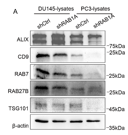
Journal: Frontiers In Immunology
アプリケーション: WB
交差性: Human
掲載日: 2025 Sept
-
Citation
-
Helicobacter pylori induced miR-362-5p upregulation drives gastric cancer progression and links hepatocellular carcinoma through an exosome-dependent pathway
Author: Jianhui Zhang, Shuzhen Liu, Juan Zhang, Mingzhu Feng, Shu Chen, Yinuo Zhang, Zekun Sun, Xinying Cao, Chao Gao, Xiaofei Ji, Huilin Zhao
PMID: 40406521
Journal: Frontiers In Cellular And Infection Microbiology
アプリケーション: WB
交差性: Human
掲載日: 2025 May
-
Citation
-
Quercetin-loaded exosomes delivery system prevents myopia progression by targeting endoplasmic reticulum stress and ferroptosis in scleral fibroblasts
Author: Lianghui Zhao, Xiaoyun Dong, Bin Guo, Jike Song, Hongsheng Bi
PMID: 40520556
Journal: Materials Today Bio
アプリケーション: WB
交差性: Human
掲載日: 2025 May
-
Citation
-
Apoptotic Vesicles Derived from Mesenchymal Stem Cells Ameliorate Hypersensitivity Responses via Inducing CD8+ T Cells Apoptosis with Calcium Overload and Mitochondrial Dysfunction
Author: Anqi Liu, Peng Peng, Changze Wei, Fanhui Meng, Xiaoyao Huang, Peisheng Liu, Siyuan Fan, Xinyue Cai, Meiling Wu, Zilin Xuan, Qing Liu, Xinyu Qiu, Zhenlai Zhu, Hao Guo
PMID: 40089865
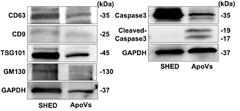
Journal: Advanced Science
アプリケーション: WB
交差性: Human
掲載日: 2025 Mar
-
Citation
-
Curcumin-loaded milk-derived sEVs fused with platelet membrane attenuate endothelial senescence and promote spinal cord injury recovery in diabetic mice
Author: Yaozhi He, Siyuan Zou, Jiawei Wang, Wenbin Zhang, Sheng Lu, Mengxian Jia, Yumin Wu, Xiaowu Lin, Ziwei Fan, Qishun Liang, Yizhe Sheng, Qichuan Zhuge, Bi Chen, Minyu Zhu, Honglin Teng
PMID: 40688662
Journal: Materials Today Bio
アプリケーション: WB
交差性: Human
掲載日: 2025 Jun
-
Citation
-
Extracellular vesicle therapy for acute pancreatitis: experimental validation of mesenchymal stem cell-derived nanovesicles
Author: Wu Yue, Liu Yan, Liu Yiping, Liu Zhiling, Yao Jiaqi, Wen Qingping
PMID: 40713893
Journal: BMC Pharmacology & Toxicology
アプリケーション: WB
交差性: Human
掲載日: 2025 Jul
-
Citation
-
Deciphering the lipid profile: A quantitative lipidomic investigation into extracellular vesicles derived from human, ewe, and goat colostrum
Author: Yue Jiang, Junru Zhu, Jiaxin Liu, Haoyuan Zhang, Pei Zhang, Lei Zhang, Jinxing Hou, Peishuai Tong, Zengkai Li, Jianhua Zhao, Xiaopeng An, Yuxuan Song
PMID: 40675480
Journal: Journal Of Dairy Science
アプリケーション: WB
交差性: Goat,Human
掲載日: 2025 Jul
-
Citation
-
Liver-Secreted Extracellular Vesicles Promote Cirrhosis-Associated Skeletal Muscle Injury Through mtDNA-cGAS/STING Axis
Author: Xiaoli Fan, Yunke Peng, Bo Li, Xiaoze Wang, Yifeng Liu, Yi Shen, Guofeng Liu, Yanyi Zheng, Qiaoyu Deng, Jingping Liu, Li Yang
PMID: 39804962
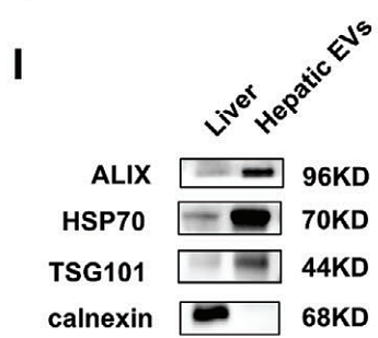
Journal: Advanced Science
アプリケーション: WB
交差性: Mouse
掲載日: 2025 Jan
-
Citation
-
The trojan horse strategy in T-ALL therapy by engineering T-ALL-derived exosomes for targeted delivery of Isoliquiritigenin to the bone marrow to conquer Drug-Resistant T-ALL in PDX model
Author: Yong Liu, Lindi Li, Cheng Ouyang, Zefan Du, Su Liu, Hailin Zou, Chunmou Li, Junbin Huang, Yucai Cheng, Mengyao Tian, Tianwen Li, Jiani Mo, Yujiang Chen, Mo Yang, Hui Chao, Jun Wu, Chun Chen
PMID: 3NOPMID25031003
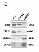
Journal: Chemical Engineering Journal
アプリケーション: WB
交差性: Human
掲載日: 2025 Jan
-
Citation
-
Emodin Alleviates Acute Pancreatitis-Associated Acute Lung Injury by Inhibiting Serum Exosomal miRNA-21-3p-Induced M1 Alveolar Macrophage Polarisation
Author: Bowen Lan, Xuanchi Dong, Qi Yang, Haiyun Wen, Yibo Zhang, Fan Li, Yinan Cao, Zhe Chen, Hailong Chen
PMID: 40845083
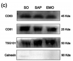
Journal: Journal Of Cellular And Molecular Medicine
アプリケーション: WB
交差性: Rat
掲載日: 2025 Aug
-
Citation
-
Targeting tumor stroma via ultrasound-activated nanodroplets: Disrupting exosome-driven microenvironment crosstalk for enhanced antitumor efficacy
Author: Yuanyuan Yang, Rui Liu, Hongtao Lv, Yading Zhao, Xiaoxuan Wang, Zijie Wang, Jie Li, Dandan Shi
PMID: 40783063
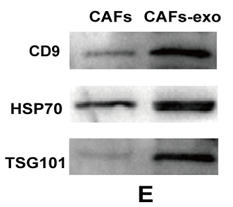
Journal: Journal Of Controlled Release
アプリケーション: WB
交差性: Mouse
掲載日: 2025 Aug
-
Citation
-
The MiR-21a-5p/transforming growth factor beta/Smad Pathway: A Potential Mechanism Underlying Effects of Qi States on PostMyocardial Infarction Heart Failure and Fibrosis
Author: Xu Lu-Hua, Wen Deng-Deng, Qi Yu-Wen, Fang Jie-Ni, Chen Ze-Tao, Zhong Da-Yuan, Feng Ting-Yu, Zeng Zhi-Cong, Lin Feng-Xia
PMID: 13NOPMID25092801
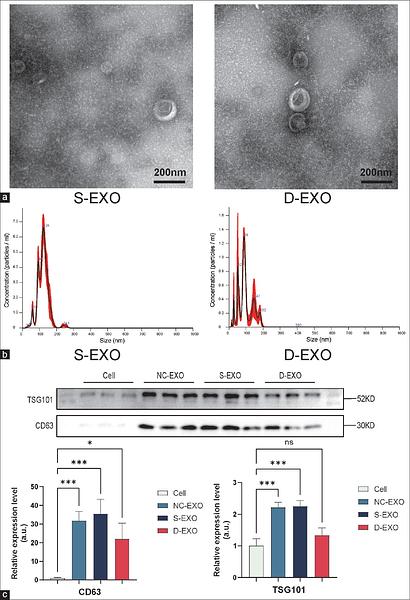
Journal: World Journal of Traditional Chinese Medicine
アプリケーション: WB
交差性: Mouse
掲載日: 2025 Aug
-
Citation
-
Exosomes Extracted from Human Umbilical Cord MSCs Contribute to Osteoarthritic Cartilage and Chondrocytes Repair Through Enhancing Autophagy While Suppressing the Wnt/β-Catenin Pathway
Author: Qin Shangzhu, Zhang Aijie, Duan Lian, Lin Fang, Zhao Mingcai
PMID: 40232653
Journal: Tissue Engineering And Regenerative Medicine
アプリケーション: WB
交差性: Human
掲載日: 2025 Apr
-
Citation
-
Mechanism of Tau Protein Incorporation into Exosomes via Cooperative Recognition of KFERQ-like Motifs by LAMP2A and HSP70
Author: Shan Xu, Kangyan Liu, Shiyan Qian, Jingying Wu, Jialing Hu, Dongming Zhou, Tingting Zheng
PMID: 40187566
Journal: Neurochemistry International
アプリケーション: WB
交差性: Human
掲載日: 2025 Apr
-
Citation
-
Single-step negative isolation and concentration of extracellular vesicles by graphene oxide composite hydrogels
Author: Qi Yang, Xinxin Liu, Kaiguang Yang, Peng Ge, Bowen Lan, Zhigang Sui, Yu Liang, Guixin Zhang, Hailong Chen, Huiming Yuan, Lihua Zhang
PMID: 11NOPMID25060302
Journal: Chemical Engineering Journal
アプリケーション: WB
交差性: Rat
掲載日: 2025 Apr
-
Citation
-
Metabolic Reprogramming of CD4+ T Cells by Mesenchymal Stem Cell-Derived Extracellular Vesicles Attenuates Autoimmune Hepatitis Through Mitochondrial Protein Transfer
Author: Mengyi Shen, Leyu Zhou, Xiaoli Fan, Ruiqi Wu, Shuyun Liu, Qiaoyu Deng, Yanyi Zheng, Jingping Liu, Li Yang
PMID: 39345912
Journal: International Journal Of Nanomedicine
アプリケーション: WB
交差性: Mouse
掲載日: 2024 Sept
-
Citation
-
Cancer-associated fibroblast-derived exosome Leptin promotes malignant biological lineage in pancreatic ductal adenocarcinoma by regulating ABL2 via miR-224-3p
Author: Zhang Li, Chen Yesheng, Dai Yihe, Mou Weicheng, Deng Pan, Jin Yan, Xu Jing, Jin Yun
PMID: 39298063

Journal: Molecular Biology Reports
アプリケーション: WB
交差性: Human
掲載日: 2024 Sept
-
Citation
-
Engineering extracellular vesicles derived from endothelial cells sheared by laminar flow for anti-atherosclerotic therapy through reprogramming macrophage
Author: Chunli Li ,et al
PMID: 39270628
Journal: Biomaterials
アプリケーション: WB
交差性: Human
掲載日: 2024 Sep
-
Citation
-
Phosphatidylserine-mediated uptake of extracellular vesicles by hepatocytes ameliorates liver ischemia-reperfusion injury
Author: Rongrong Li,et al
PMID: 39397123
Journal: Apoptosis
アプリケーション: WB
交差性: Mouse
掲載日: 2024 Oct
-
Citation
-
Plasma-derived Exosomal miR-25-3p and miR-23b-3p as Predictors of Response to Chemoradiotherapy in Esophageal Squamous Cell Carcinoma
Author: Zhao Jing,et al
PMID: 39380461
Journal: Technology In Cancer Research & Treatment
アプリケーション:
交差性:
掲載日: 2024 Oct
-
Citation
-
Matrix vesicles from osteoblasts promote atherosclerotic calcification
Author: Xiaoli Wang,et al
PMID: 39580186
Journal: Matrix Biology
アプリケーション: WB
交差性: Human
掲載日: 2024 Oct
-
Citation
-
Serum starvation induces cytosolic DNA trafficking via exosome and autophagy-lysosome pathway in microglia
Author: Zhou Liyan,et al
PMID: 39880132
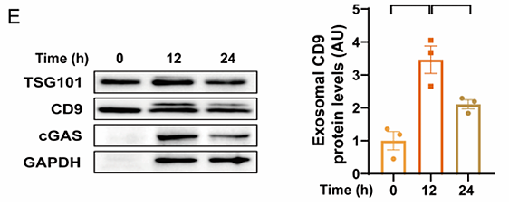
Journal: Preprint And Has Not Been Certified By Peer Review
アプリケーション: WB
交差性: Mouse
掲載日: 2024 May
-
Citation
-
Investigating the therapeutic effects and mechanisms of Carthamus tinctorius L.-derived nanovesicles in atherosclerosis treatment
Author: Yang Rongfeng,et al
PMID: 38475787
Journal: Cell Communication And Signaling
アプリケーション: WB
交差性: Mouse
掲載日: 2024 Mar
-
Citation
-
Mesenchymal stem cells-derived exosomes alleviate temporomandibular joint disc degeneration in temporomandibular joint disorder
Author: Yang Yutao,et al
PMID: 38936248
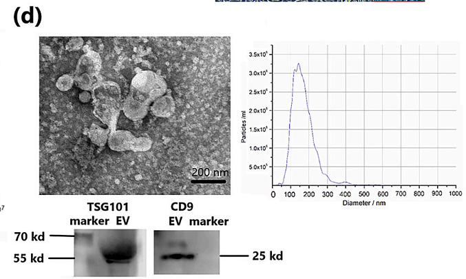
Journal: Biochemical And Biophysical Research Communications
アプリケーション: WB
交差性: Rat
掲載日: 2024 Jun
-
Citation
-
iMSC exosome delivers hsa-mir-125b-5p and strengthens acidosis resilience through suppression of Asic1 protein in cerebral ischemia-reperfusion
Author: Dong Kai,et al
PMID: 39019215
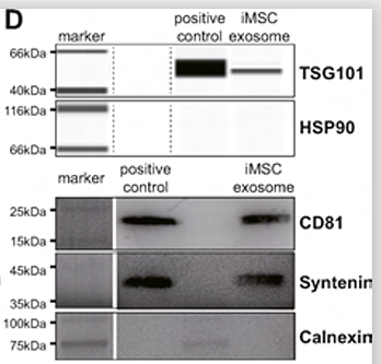
Journal: Journal Of Biological Chemistry
アプリケーション: WB
交差性: Mouse
掲載日: 2024 Jul
-
Citation
-
Nintedanib-loaded exosomes from adipose-derived stem cells inhibit pulmonary fibrosis induced by bleomycin
Author: Cai Liyun, Wang Jie, Yi Xue, Yu Shuwei, Wang Chong, Zhang Liyuan, Zhang Xiaoling, Cheng Lixian, Ruan Wenwen, Dong Feige, Su Ping, Shi Ying
PMID: 38245633
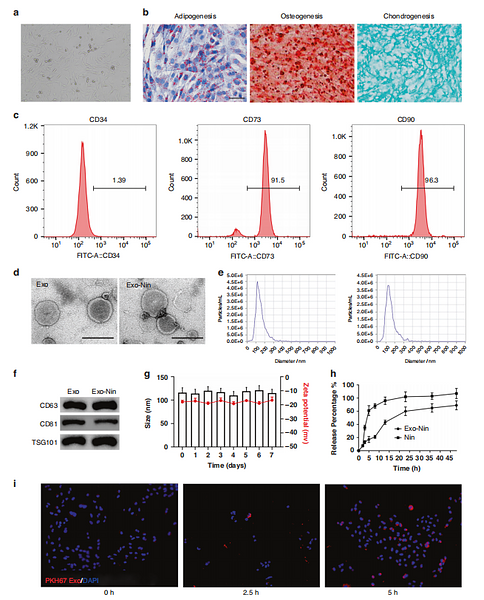
Journal: Pediatric Research
アプリケーション: WB
交差性: Mouse
掲載日: 2024 Jan
-
Citation
-
miR-21-loaded BMSC-derived Exosomes repair ovarian function of autoimmune premature ovarian insufficiency by targeting MSX1
Author: Yang Yutao,et al
PMID: 38582043
Journal: Reproductive Biomedicine Online
アプリケーション: WB
交差性: Mouse
掲載日: 2024 Jan
-
Citation
-
Designed Directional Growth of Ti-Metal-Organic Frameworks for Decoding Alzheimer's Disease-Specific Exosome Metabolites
Author: Chen Yijie,et al
PMID: 38300748
Journal: Analytical Chemistry
アプリケーション: WB
交差性: Human
掲載日: 2024 Feb
-
Citation
-
miR-21-loaded bone marrow mesenchymal stem cell-derived exosomes inhibit pyroptosis by targeting MALT1 to repair chemotherapy-induced premature ovarian insufficiency
Author: Tang Lichao, Yang Yutao, Yang Mingxin, Xie Jiaxin, Zhuo Aiping, Wu Yanhong, Mao Mengli, Zheng Youhong, Fu Xiafei
PMID: 39707056
Journal: Cell Biology And Toxicology
アプリケーション: WB
交差性: Mouse
掲載日: 2024 Dec
-
Citation
-
Engaging natural regulatory myeloid cells to restrict T-cell hyperactivation-induced liver inflammation via extracellular vesicle-mediated purine metabolism regulation
Author: Yang Fan,et al
PMID: 39239508
Journal: Theranostics
アプリケーション: WB
交差性: Mouse
掲載日: 2024 Aug
-
Citation
-
Adipose-Derived Mesenchymal Stem Cell-Derived Exosomes Biopotentiated Extracellular Matrix Hydrogels Accelerate Diabetic Wound Healing and Skin Regeneration
Author: Yanling Song, Yuchan You, Xinyi Xu, Jingyi Lu, Xiajie Huang, Jucong Zhang, Luwen Zhu, Jiahao Hu, Xiaochuan Wu, Xiaoling Xu, Weiqiang Tan, Yongzhong Du
PMID: 37712174
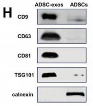
Journal: Advanced Science
アプリケーション: WB
交差性: Mouse
掲載日: 2023 Sept
-
Citation
-
Metabolic profiling of urinary exosomes for systemic lupus erythematosus discrimination based on HPL-SEC/MALDI-TOF MS
Author: Yan S, Huang Z, Chen X, et al
PMID: 37644324
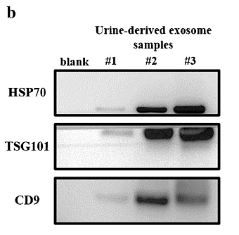
Journal: Analytical And Bioanalytical Chemistry
アプリケーション: WB
交差性: Human
掲載日: 2023 Nov
-
Citation
-
Emodin Ameliorates Severe Acute Pancreatitis-Associated Acute Lung Injury in Rats by Modulating Exosome-Specific miRNA Expression Profiles
Author: Qi Yang, Yalan Luo, Peng Ge, Bowen Lan, Jin Liu, Haiyun Wen, Yinan Cao, Zhenxuan Sun, Guixin Zhang, Huiming Yuan, Lihua Zhang, Hailong Chen
PMID: 38026528
Journal: International Journal Of Nanomedicine
アプリケーション: WB
交差性: Rat
掲載日: 2023 Nov
-
Citation
-
The Cellular Mechanism of Acupuncture for Ulcerative Colitis based on the Communication of Telocytes
Author: Bai Xuebing, Mei Lu, Shi Yonghong, Huang Haixiang, Guo Yanna, Liang Chunhua, Yang Min, Wu Ruizhi, Zhang Yingxin, Chen Qiusheng
PMID: 37749671
Journal: Microscopy And Microanalysis
アプリケーション: IF,IF-cell
交差性: Rabbit
掲載日: 2023 Mar
-
Citation
-
Embryonic stem cell-derived extracellular vesicles rejuvenate senescent cells and antagonize aging in mice
Author:
PMID: 37449253
Journal: Bioactive Materials
アプリケーション: WB
交差性: Mouse
掲載日: 2023 Jul
-
Citation
-
A protocol of carbonized on-column enrichment for urinary exosomal N-glycans profiling
Author:
PMID: 36592588
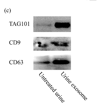
Journal: Journal Of Chromatography B-Analytical Technologies In The Biomedical And Life Sciences
アプリケーション: WB
交差性: Human
掲載日: 2023 Jan
-
Citation
-
D-mannose facilitates immunotherapy and radiotherapy of triple-negative breast cancer via degradation of PD-L1
Author: Zhang, R., Yang, Y., Dong, W., Lin, M., He, J., Zhang, X., Tian, T., Yang, Y., Chen, K., Lei, Q. Y., Zhang, S., Xu, Y., & Lv, L.
PMID: 35181605
Journal: Proceedings Of The National Academy Of Sciences Of The United States Of America
アプリケーション: WB
交差性: Human
掲載日: 2022 Feb
-
Citation
-
Myeloid-derived suppressor cells ameliorate liver mitochondrial damage to protect against autoimmune hepatitis by releasing small extracellular vesicles
Author: Shen, M., Fan, X., Shen, Y., Wang, X., Wu, R., Wang, Y., Huang, C., Zhao, S., Zheng, Y., Men, R., Luo, X., & Yang, L.
PMID: 36516541
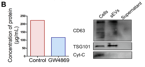
Journal: International Immunopharmacology
アプリケーション: WB
交差性: Human
掲載日: 2022 Dec
-
Citation
-
Embryonic stem cell-derived extracellular vesicles promote the recovery of kidney injury
Author:
PMID: 34215331
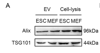
Journal: Stem Cell Research & Therapy
アプリケーション: WB
交差性: Mouse
掲載日: 2021 Jul
-
Citation
-
In vivo multivesicular bodies and their exosomes in the absorptive cells of the zebrafish (Danio Rerio) gut
Author: Qiusheng Chen
PMID: 30885742
Journal: Fish & Shellfish Immunology
アプリケーション: IHC-P,IF
交差性: zebrafish
掲載日: 2019 May
-
Citation
-
In Vivo Multivesicular Body and Exosome Secretion in the Intestinal Epithelial Cells of Turtles During Hibernation.
Author: Qiusheng Chen
PMID: 31656212
Journal: Microscopy And Microanalysis
アプリケーション: IHC-P,IF
交差性: Turtle
掲載日: 2019 Dec
-
Citation

