CD31 Recombinant Rabbit Monoclonal Antibody [SU03-59]
製品仕様
今すぐ注文,【现货】
問い合わせをクリックCatalog# ET1608-48
CD31 Recombinant Rabbit Monoclonal Antibody [SU03-59]
-
WB
-
IHC-P
-
FC
-
IP
-
IF-Cell
-
IF-Tissue
-
Human
-
HA750151
不含抗保成分
-
ET1608-48
含抗保成分
-
unconjugated
概要
製品名
CD31 Recombinant Rabbit Monoclonal Antibody [SU03-59]
抗体の種類
Recombinant Rabbit monoclonal Antibody
免疫原
Synthetic peptide within Human CD31 aa 689-738 / 738.
種属反応性
Human
検証された応用例
WB, IHC-P, FC, IP, IF-Cell, IF-Tissue
分子量
Predicted band size: 83 kDa
陽性対照
THP-1 cell lysate, human tonsil tissue, THP-1.
結合
unconjugated
クローン番号
SU03-59
RRID
製品の特徴
形態
Liquid
保存方法
Shipped at 4℃. Store at +4℃ short term (1-2 weeks). It is recommended to aliquot into single-use upon delivery. Store at -20℃ long term.
保存用バッファー
1*TBS (pH7.4), 0.05% BSA, 40% Glycerol. Preservative: 0.05% Sodium Azide.
アイソタイプ
IgG
精製方法
Protein A affinity purified.
応用希釈度
-
WB
-
1:1,000-1:2,000
-
IHC-P
-
1:50-1:1,000
-
FC
-
1:50-1:100
-
IP
-
1-2μg/sample
-
IF-Cell
-
1:100
-
IF-Tissue
-
1:200
論文における応用例
| IF | 確認する 5 以下の論文 |
| WB | 確認する 3 以下の論文 |
| IHC-P | 確認する 3 以下の論文 |
| IF-tissue | 確認する 1 以下の論文 |
| IHC | 確認する 1 以下の論文 |
論文における種属
| Human | 確認する 7 以下の論文 |
| Mouse | 確認する 6 以下の論文 |
| Rat | 確認する 1 以下の論文 |
ターゲット
機能
PECAM-1 is a cell-cell adhesion protein which interacts with other PECAM-1 molecules through homophilic interactions or with non-PECAM-1 molecules through heterophilic interactions. Homophilic interactions between PECAM-1 molecules are mediated by antiparallel interactions between extracellular Ig-like domain 1 and Ig-like domain 2. These interactions are regulated by the level of PECAM-1 expression. Homophilic interactions occur, only when the surface expression of PECAM-1 is high. Otherwise, when expression is low, heterophilic interactions occur.
背景文献
1. Doi H et al. Potency of umbilical cord blood- and Wharton\'s jelly-derived mesenchymal stem cells for scarless wound healing. Sci Rep 6:18844 (2016).
2. Yang Y et al. The Increased Expression of Connexin and VEGF in Mouse Ovarian Tissue Vitrification by Follicle Stimulating Hormone. Biomed Res Int 2015:397264 (2015).
組織特異性
Expressed on platelets and leukocytes and is primarily concentrated at the borders between endothelial cells. Expressed in human umbilical vein endothelial cells (HUVECs) (at protein level). Expressed on neutrophils (at protein level). Isoform Long predominates in all tissues examined. Isoform Delta12 is detected only in trachea. Isoform Delta14-15 is only detected in lung. Isoform Delta14 is detected in all tissues examined with the strongest expression in heart. Isoform Delta15 is expressed in brain, testis, ovary, cell surface of platelets, human umbilical vein endothelial cells (HUVECs), Jurkat T-cell leukemia, human erythroleukemia (HEL) and U-937 histiocytic lymphoma cell lines (at protein level).
翻訳後修飾
Phosphorylated on Ser and Tyr residues after cellular activation by src kinases. Upon activation, phosphorylated on Ser-729 which probably initiates the dissociation of the membrane-interaction segment (residues 709-729) from the cell membrane allowing the sequential phosphorylation of Tyr-713 and Tyr-690. Constitutively phosphorylated on Ser-734 in resting platelets. Phosphorylated on tyrosine residues by FER and FES in response to FCER1 activation (By similarity). In endothelial cells Fyn mediates mechanical-force (stretch or pull) induced tyrosine phosphorylation.; Palmitoylation by ZDHHC21 is necessary for cell surface expression in endothelial cells and enrichment in membrane rafts.
亜細胞局在
Cell junction. Cell membrane. Membrane.
UNIPROT #
別名
Adhesion molecule antibody
CD31 antibody
CD31 antigen antibody
CD31 EndoCAM antibody
EndoCAM antibody
FLJ34100 antibody
FLJ58394 antibody
GPIIA antibody
GPIIA' antibody
PECA1 antibody
展開Adhesion molecule antibody
CD31 antibody
CD31 antigen antibody
CD31 EndoCAM antibody
EndoCAM antibody
FLJ34100 antibody
FLJ58394 antibody
GPIIA antibody
GPIIA' antibody
PECA1 antibody
PECA1_HUMAN antibody
Pecam 1 antibody
PECAM 1 CD31 EndoCAM antibody
PECAM antibody
PECAM-1 antibody
Pecam1 antibody
Platelet and endothelial cell adhesion molecule 1 antibody
Platelet endothelial cell adhesion molecule antibody
Platelet/endothelial cell adhesion molecule 1 antibody
折りたたむ画像
-

Western blot analysis of CD31 on THP-1 cell lysates with Rabbit anti-CD31 antibody (ET1608-48) at 1/2,000 dilution.
Lysates/proteins at 15 µg/Lane.
Predicted band size: 83 kDa
Observed band size: 130 kDa
Exposure time: 20 seconds;
4-20% SDS-PAGE gel.
Proteins were transferred to a PVDF membrane and blocked with 5% NFDM/TBST for 1 hour at room temperature. The primary antibody (ET1608-48) at 1/2,000 dilution was used in 5% NFDM/TBST at room temperature for 2 hours. Goat Anti-Rabbit IgG - HRP Secondary Antibody (HA1001) at 1:50,000 dilution was used for 1 hour at room temperature. -

CD31 was immunoprecipitated in 0.2mg THP-1 cell lysate with ET1608-48 at 2 µg/10 µl beads. Western blot was performed from the immunoprecipitate using ET1608-48 at 1/1,000 dilution. Anti-Rabbit IgG for IP Nano-secondary antibody (NBI01H) at 1/5,000 dilution was used for 1 hour at room temperature.
Lane 1: THP-1 cell lysate (input)
Lane 2: Rabbit IgG instead of ET1608-48 in THP-1 cell lysate
Lane 3: ET1608-48 IP in THP-1 cell lysate
Blocking/Dilution buffer: 5% NFDM/TBST
Exposure time: 5 seconds -

Immunohistochemical analysis of paraffin-embedded human tonsil tissue with Rabbit anti-CD31 antibody (ET1608-48) at 1/1,000 dilution.
The section was pre-treated using heat mediated antigen retrieval with Tris-EDTA buffer (pH 9.0) for 20 minutes. The tissues were blocked in 1% BSA for 20 minutes at room temperature, washed with ddH2O and PBS, and then probed with the primary antibody (ET1608-48) at 1/1,000 dilution for 1 hour at room temperature. The detection was performed using an HRP conjugated compact polymer system. DAB was used as the chromogen. Tissues were counterstained with hematoxylin and mounted with DPX. -

Flow cytometric analysis of CD31 was done on THP-1 cells. The cells were fixed, permeabilized and stained with the primary antibody (ET1608-48, 1/50) (blue). After incubation of the primary antibody at room temperature for an hour, the cells were stained with a Alexa Fluor 488-conjugated Goat anti-Rabbit IgG Secondary antibody at 1/1,000 dilution for 30 minutes.Unlabelled sample was used as a control (cells without incubation with primary antibody; red).
-

Immunocytochemistry analysis of THP-1 cells labeling CD31 with Rabbit anti-CD31 antibody (ET1608-48) at 1/100 dilution.
Cells were fixed in 4% paraformaldehyde for 20 minutes at room temperature, permeabilized with 0.1% Triton X-100 in PBS for 5 minutes at room temperature, then blocked with 1% BSA in 10% negative goat serum for 1 hour at room temperature. Cells were then incubated with Rabbit anti-CD31 antibody (ET1608-48) at 1/100 dilution in 1% BSA in PBST overnight at 4 ℃. Goat Anti-Rabbit IgG H&L (iFluor™ 488, HA1121) was used as the secondary antibody at 1/1,000 dilution. PBS instead of the primary antibody was used as the secondary antibody only control. Nuclear DNA was labelled in blue with DAPI.
Beta tubulin (M1305-2, red) was stained at 1/100 dilution overnight at +4℃. Goat Anti-Mouse IgG H&L (iFluor™ 594, HA1126) was used as the secondary antibody at 1/1,000 dilution. -

Immunofluorescence analysis of paraffin-embedded human tonsil tissue labeling CD31 with Rabbit anti-CD31 antibody (ET1608-48) at 1/200 dilution.
The section was pre-treated using heat mediated antigen retrieval with Tris-EDTA buffer (pH 9.0) for 20 minutes. The tissues were blocked in 10% negative goat serum for 1 hour at room temperature, washed with PBS, and then probed with the primary antibody (ET1608-48, green) at 1/200 dilution overnight at 4 ℃, washed with PBS. Goat Anti-Rabbit IgG H&L (iFluor™ 488, HA1121) was used as the secondary antibody at 1/1,000 dilution. Nuclei were counterstained with DAPI (blue).
ご注意ください: All products are "FOR RESEARCH USE ONLY AND ARE NOT INTENDED FOR DIAGNOSTIC OR THERAPEUTIC USE"
論文での実績
-
Astragaloside IV Binds with RhoA, Inhibits EndMT and Ameliorates Myocardial Fibrosis in Mice
Author: Xuan Liu
PMID: 40916081
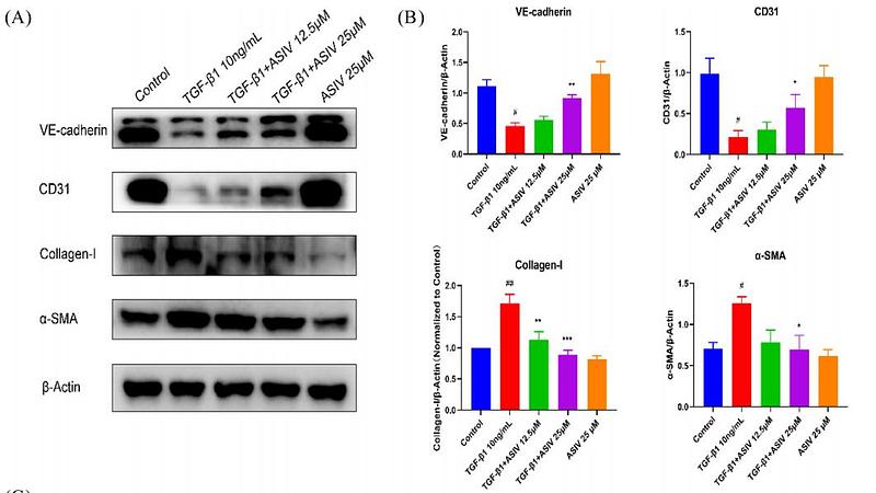
Journal: American Journal Of Chinese Medicine
アプリケーション: WB
交差性: Human
掲載日: 2025 Sept
-
Citation
-
Decellularized ECM from Blastema Krt42⁺ Cells Promotes Skin Regeneration via Immunometabolic Modulation of Macrophages
Author: Nian Zhang, Liru Hu, Chengzhi Zhao, Zhiwei Cao, Xian Liu, Jian Pan
PMID: 40442963
Journal: Small
アプリケーション: IHC
交差性: Mouse
掲載日: 2025 May
-
Citation
-
Urokinase-loaded Pt Quantum Dot Self-assembly Nanoparticle for Inflammation Elimination and Fibrinolytic Thrombus Therapy
Author: Zhaoxi Peng, Yu Cao, Hongji Pu, Cheng Cao, Wenxin Yang, Sen Yang, Yijun Liu, Peng Qiu, Xinrui Yang, Ruihua Wang, Chaowen Yu, Haoqi Liu, Kaichuang Ye, Xinwu Lu
PMID: 16NOPMID25050801
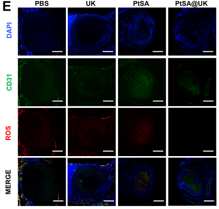
Journal: Materials Chemistry Frontiers
アプリケーション: IF
交差性: Mouse
掲載日: 2025 Mar
-
Citation
-
Injectable Scaffolds with Hierarchically Porous Structure and Augment Paracrine Activity for Minimally Invasive Precision Medicine
Author: Lin Du, Hongjian Zhang, Ziyi Zhao, Xueru Ma, Jimin Huang, Jinzhou Huang, Chengtie Wu
PMID: 40620145
Journal: Materials Horizons
アプリケーション: IF
交差性: Rat,Human
掲載日: 2025 Jun
-
Citation
-
Magnetic Nanoactuator-Protein Fiber Coated Hydrogel Dressing for Well-Balanced Skin Wound Healing and Tissue Regeneration
Author: Chenlong He, Ming Yin, Han Zhou, Jingwen Qin, Shengming Wu, Huawei Liu, Xiaoyu Yu, Jing Chen, Hongyi Zhang, Lin Zhang, Yilong Wang
PMID: 39749690

Journal: ACS Applied Nano Materials
アプリケーション: IF
交差性: Mouse
掲載日: 2025 Jan
-
Citation
-
Controlled Release of Cold Atmospheric Plasma by Gelatin Scaffold Enhances Wound Healing via Macrophage Modulation
Author: Nian Zhang, Guang Yang, Yan Wu, Liru Hu, Chengzhi Zhao, Hang-Hang Liu, Li Wu, Jian Pan, Xian Liu
PMID: 40013441
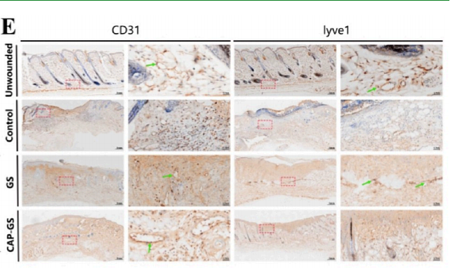
Journal: ACS Applied Materials & Interfaces
アプリケーション: IHC-P
交差性: Mouse
掲載日: 2025 Feb
-
Citation
-
Polyphenol-Based Photothermal Nanoparticles with Sprayable Capability for Self-regulation of Microenvironment to Accelerate Diabetic Wound Healing
Author: Huang Xiuhong,Fu Meimei,Lu Min,et al
PMID: NO PMID202406
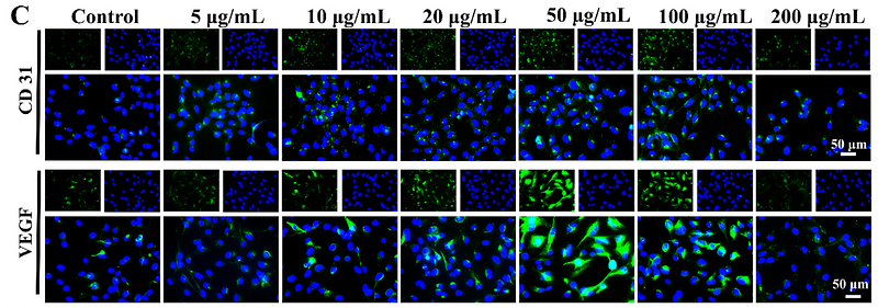
Journal: Engineered Regeneration
アプリケーション: IF
交差性: Human
掲載日: 2024 Jun
-
Citation
-
Decoding the Cell Atlas and Inflammatory Features of Human Intracranial Aneurysm Wall by Single‐Cell RNA Sequencing
Author: Hang Ji, Yue Li, Haogeng Sun, Ruiqi Chen, Ran Zhou, Yongbo Yang, Rong Wang, Chao You, Anqi Xiao, Liu Yi
PMID: 38390814
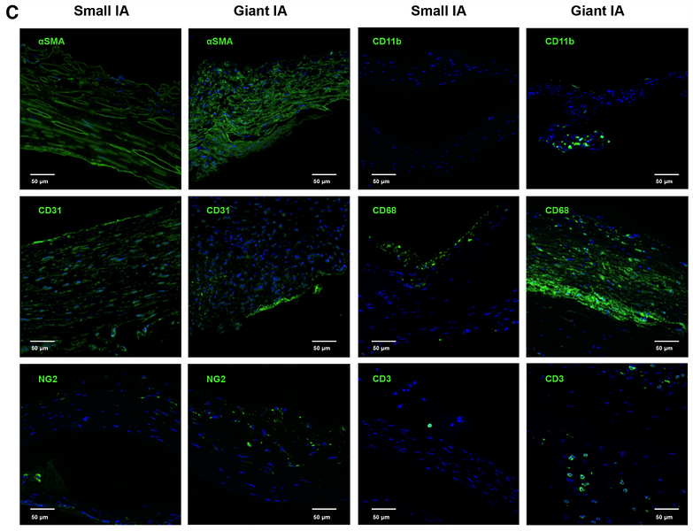
Journal: Journal Of The American Heart Association
アプリケーション: IF
交差性: Human
掲載日: 2024 Feb
-
Citation
-
N6-methyladenosine-modified TRAF1 promotes sunitinib resistance by regulating apoptosis and angiogenesis in a METTL14-dependent manner in renal cell carcinoma
Author: Chen, Y., Lu, Z., Qi, C., Yu, C., Li, Y., Huan, W., Wang, R., Luo, W., Shen, D., Ding, L., Ren, L., Xie, H., Xue, D., Wang, M., Ni, K., Xia, L., Qian, J., & Li, G.
PMID: 35538475
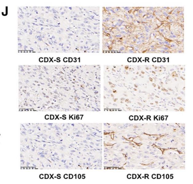
Journal: Molecular Cancer
アプリケーション: IHC-P
交差性: Human
掲載日: 2022 May
-
Citation
-
Yes-associated protein promotes endothelial-to-mesenchymal transition of endothelial cells in choroidal neovascularization fibrosis
Author:
PMID: 35601164
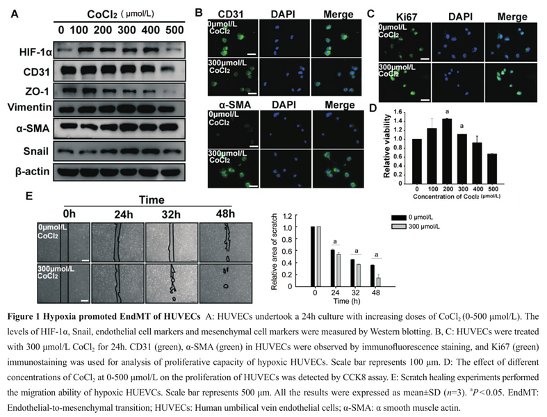
Journal: International Journal Of Ophthalmology
アプリケーション: WB
交差性: Human
掲載日: 2022 May
-
Citation
-
Microglia Regulate Blood–Brain Barrier Integrity via MiR‐126a‐5p/MMP9 Axis during Inflammatory Demyelination
Author:
PMID: 35758549
Journal: Advanced Science
アプリケーション: IF-tissue
交差性: Mouse
掲載日: 2022 Aug
-
Citation
-
A single-cell atlas of liver metastases of colorectal cancer reveals reprogramming of the tumor microenvironment in response to preoperative chemotherapy
Author: Che, L. H., Liu, J. W., Huo, J. P., Luo, R., Xu, R. M., He, C., Li, Y. Q., Zhou, A. J., Huang, P., Chen, Y. Y., Ni, W., Zhou, Y. X., Liu, Y. Y., Li, H. Y., Zhou, R., Mo, H., & Li, J. M.
PMID: 34489408
Journal: Cell Discovery
アプリケーション: IHC-P
交差性: Human
掲載日: 2021 Sep
-
Citation
-
Targeting Tumor Hypoxia with Stimulus-Responsive Nanocarriers in Overcoming Drug Resistance and Monitoring Anticancer Efficacy
Author: Min Han,JianQing Gao
PMID: 29545193
Journal: Acta Biomaterialia
アプリケーション: WB
交差性: Mouse
掲載日: 2018 Apr
-
Citation
同じターゲット & 同じ経路の製品
CD31 Recombinant Antibody [7-A1-R] - Rabbit IgG (Chimeric) - BSA and Azide free
Application: WB,IF-Cell,IHC-P,FC
Reactivity: Human
Conjugate: unconjugated
CD31 Recombinant Antibody [7-A1-R] - Rabbit IgG (Chimeric)
Application: WB,IF-Cell,IHC-P,FC,mIHC
Reactivity: Human
Conjugate: unconjugated
CD31 Recombinant Rabbit Monoclonal Antibody [SU03-59] - BSA and Azide free
Application: WB,IHC-P,FC,IP,IF-Cell,IF-Tissue
Reactivity: Human
Conjugate: unconjugated
CD31 Recombinant Antibody - Rabbit IgG (Chimeric)
Application: mIHC
Reactivity: Human
Conjugate: unconjugated
CD31 Recombinant Antibody [7-A1-R] - Rat IgG1 (Chimeric) - BSA and Azide free
Application: IHC-P
Reactivity: Human
Conjugate: unconjugated
CD31 Recombinant Mouse Monoclonal Antibody [7-A1-R] - BSA and Azide free
Application: WB,IF-Cell,IHC-P
Reactivity: Human
Conjugate: unconjugated
CD31 Mouse Monoclonal Antibody
Application: mIHC
Reactivity: Human
Conjugate: unconjugated
CD31 Recombinant Antibody [7-A1-R] - Rat IgG1 (Chimeric)
Application: IHC-P
Reactivity: Human
Conjugate: unconjugated
CD31 Rabbit Polyclonal Antibody
Application: WB,IF-Cell,FC,IHC-P
Reactivity: Human,Mouse,Rat
Conjugate: unconjugated
iFluor™ 488 Conjugated CD31 Recombinant Rabbit Monoclonal Antibody [SU03-59]
Application: IF-Tissue
Reactivity: Human
Conjugate: iFluor™ 488
CD31 Mouse Monoclonal Antibody [7-A1]
Application: WB,IF-Cell,IHC-P,mIHC
Reactivity: Human
Conjugate: unconjugated
CD31 Recombinant Mouse Monoclonal Antibody [7-A1-R]
Application: WB,IF-Cell,IHC-P
Reactivity: Human
Conjugate: unconjugated


