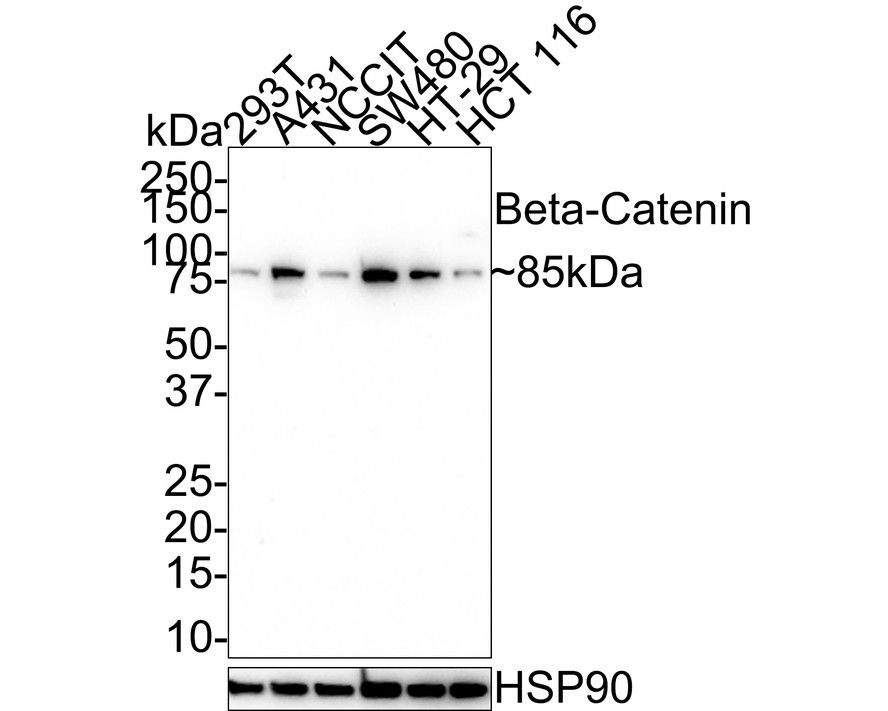
Beta Catenin Rabbit Polyclonal Antibody
製品仕様
今すぐ注文,【现货】
問い合わせをクリックCatalog# 0407-16
Beta Catenin Rabbit Polyclonal Antibody
-
WB
-
IF-Cell
-
Human
-
Mouse
-
Rat
-
Zebrafish
-
unconjugated
概要
製品名
Beta Catenin Rabbit Polyclonal Antibody
抗体の種類
Rabbit Polyclonal Antibody
免疫原
Synthetic peptide within N-terminal human beta Catenin.
種属反応性
Human, Mouse, Rat, Zebrafish
検証された応用例
WB, IF-Cell
分子量
Predicted band size: 85 kDa
陽性対照
A431 cell lysate, NIH/3T3 cell lysate, C6 cell lysate, Mouse brain tissue lysate, Rat brain tissue lysate, mouse liver tissue lysates, hybrid fish (crucian-carp) heart tissue lysates, A431.
結合
unconjugated
RRID
製品の特徴
形態
Liquid
保存方法
Shipped at 4℃. Store at +4℃ short term (1-2 weeks). It is recommended to aliquot into single-use upon delivery. Store at -20℃ long term.
保存用バッファー
1*PBS (pH7.4), 0.2% BSA, 40% Glycerol. Preservative: 0.05% Sodium Azide.
アイソタイプ
IgG
精製方法
Immunogen affinity purified.
応用希釈度
-
WB
-
1:500-1:5,000
-
IF-Cell
-
1:3,000
論文における応用例
| WB | 確認する 12 以下の論文 |
| IF | 確認する 2 以下の論文 |
| IF-cell | 確認する 1 以下の論文 |
| IHC-P | 確認する 1 以下の論文 |
論文における種属
| Human | 確認する 5 以下の論文 |
| Mouse | 確認する 4 以下の論文 |
| Zebrafish | 確認する 1 以下の論文 |
| Pig | 確認する 1 以下の論文 |
| Rat | 確認する 1 以下の論文 |
| Escherichia coli | 確認する 1 以下の論文 |
| Fish | 確認する 1 以下の論文 |
ターゲット
機能
Key downstream component of the canonical Wnt signaling pathway In the absence of Wnt, forms a complex with AXIN1, AXIN2, APC, CSNK1A1 and GSK3B that promotes phosphorylation on N-terminal Ser and Thr residues and ubiquitination of CTNNB1 via BTRC and its subsequent degradation by the proteasome. In the presence of Wnt ligand, CTNNB1 is not ubiquitinated and accumulates in the nucleus, where it acts as a coactivator for transcription factors of the TCF/LEF family, leading to activate Wnt responsive genes Involved in the regulation of cell adhesion, as component of an E-cadherin:catenin adhesion complex. Acts as a negative regulator of centrosome cohesion. Involved in the CDK2/PTPN6/CTNNB1/CEACAM1 pathway of insulin internalization. Blocks anoikis of malignant kidney and intestinal epithelial cells and promotes their anchorage-independent growth by down-regulating DAPK2. Disrupts PML function and PML-NB formation by inhibiting RANBP2-mediated sumoylation of PML. Promotes neurogenesis by maintaining sympathetic neuroblasts within the cell cycle. Involved in chondrocyte differentiation via interaction with SOX9: SOX9-binding competes with the binding sites of TCF/LEF within CTNNB1, thereby inhibiting the Wnt signaling.
背景文献
1. Kikuchi A.;"Regulation of beta-catenin signaling in the Wnt pathway."; Biochem. Biophys. Res. Commun. 268:243-248(2000).
2. Dobrosotskaya I.Y., James G.L.; "MAGI-1 interacts with beta-catenin and is associated with cell-cell adhesion structures."; Biochem. Biophys. Res. Commun. 270:903-909(2000).
3. Kim J.-S., Crooks H., Dracheva T., Nishanian T.G., Singh B., Jen J., Waldman T.; "Oncogenic beta-catenin is required for bone morphogenetic protein 4 expression in human cancer cells."; Cancer Res. 62:2744-2748(2002).
配列相同性
Belongs to the beta-catenin family.
組織特異性
Expressed in cerebellar granule neurons (at protein level).
翻訳後修飾
Phosphorylation by GSK3B requires prior phosphorylation of Ser-45 by another kinase. Phosphorylation proceeds then from Thr-41 to Ser-33. Phosphorylated by NEK2. EGF stimulates tyrosine phosphorylation. Phosphorylation on Tyr-654 decreases CDH1 binding and enhances TBP binding (By similarity). Phosphorylated on Ser-33 and Ser-37 by HIPK2. This phosphorylation triggers proteasomal degradation. Phosphorylation at Ser-552 by AMPK promotes stabilizion of the protein, enhancing TCF/LEF-mediated transcription. Phosphorylation on Ser-191 and Ser-246 by CDK5. Phosphorylation by CDK2 regulates insulin internalization (By similarity). Phosphorylation by PTK6 at Tyr-64, Tyr-142, Tyr-331 and/or Tyr-333 with the predominant site at Tyr-64 is not essential for inhibition of transcriptional activity (By similarity).; Ubiquitinated by the SCF(BTRC) E3 ligase complex when phosphorylated by GSK3B, leading to its degradation (By similarity). Ubiquitinated by a E3 ubiquitin ligase complex containing UBE2D1, SIAH1, CACYBP/SIP, SKP1, APC and TBL1X, leading to its subsequent proteasomal degradation (By similarity). Ubiquitinated and degraded following interaction with SOX9 (Probable).; S-nitrosylation at Cys-619 within adherens junctions promotes VEGF-induced, NO-dependent endothelial cell permeability by disrupting interaction with E-cadherin, thus mediating disassembly adherens junctions.; O-glycosylation at Ser-23 decreases nuclear localization and transcriptional activity, and increases localization to the plasma membrane and interaction with E-cadherin CDH1.; Deacetylated at Lys-49 by SIRT1.
亜細胞局在
Cell membrane, Synapse, Cell junction, Cytoplasm, Nucleus, Cytoskeleton.
別名
β Catenin
Beta catenin antibody
Beta-catenin antibody
Cadherin associated protein antibody
Catenin (cadherin associated protein), beta 1, 88kDa antibody
Catenin beta 1 antibody
Catenin beta-1 antibody
CATNB antibody
CHBCAT antibody
CTNB1_HUMAN antibody
展開β Catenin
Beta catenin antibody
Beta-catenin antibody
Cadherin associated protein antibody
Catenin (cadherin associated protein), beta 1, 88kDa antibody
Catenin beta 1 antibody
Catenin beta-1 antibody
CATNB antibody
CHBCAT antibody
CTNB1_HUMAN antibody
CTNNB antibody
CTNNB1 antibody
DKFZp686D02253 antibody
FLJ25606 antibody
FLJ37923 antibody
OTTHUMP00000162082 antibody
OTTHUMP00000165222 antibody
OTTHUMP00000165223 antibody
OTTHUMP00000209288 antibody
OTTHUMP00000209289 antibody
折りたたむ画像
-

Western blot analysis of Beta Catenin on different lysates with Rabbit anti-Beta Catenin antibody (0407-16) at 1/1,000 dilution.
Lane 1: A431 cell lysate (20 µg/Lane)
Lane 2: NIH/3T3 cell lysate (20 µg/Lane)
Lane 3: C6 cell lysate (20 µg/Lane)
Lane 4: Mouse brain tissue lysate (40 µg/Lane)
Lane 5: Rat brain tissue lysate (40 µg/Lane)
Predicted band size: 85 kDa
Observed band size: 85 kDa
Exposure time: 10 seconds; ECL: K1801;
4-20% SDS-PAGE gel.
Proteins were transferred to a PVDF membrane and blocked with 5% NFDM/TBST for 1 hour at room temperature. The primary antibody (0407-16) at 1/1,000 dilution was used in 5% NFDM/TBST at 4℃ overnight. Goat Anti-Rabbit IgG - HRP Secondary Antibody (HA1001) at 1/50,000 dilution was used for 1 hour at room temperature. -

Western blot analysis of Beta Catenin on mouse liver tissue lysates with Rabbit anti-Beta Catenin antibody (0407-16) at 1/500 dilution.
Lysates/proteins at 10 µg/Lane.
Predicted band size: 85 kDa
Observed band size: 85 kDa
Exposure time: 30 seconds;
8% SDS-PAGE gel.
Proteins were transferred to a PVDF membrane and blocked with 5% NFDM/TBST for 1 hour at room temperature. The primary antibody (0407-16) at 1/500 dilution was used in 5% NFDM/TBST at room temperature for 2 hours. Goat Anti-Rabbit IgG - HRP Secondary Antibody (HA1001) at 1:40,000 dilution was used for 1 hour at room temperature. -

Western blot analysis of beta Catenin on hybrid fish (crucian-carp) heart tissue lysates. Proteins were transferred to a PVDF membrane and blocked with 5% NFDM/TBST for 1 hour at room temperature. The primary antibody (0407-16, 1/500) was used in 5% NFDM/TBST at room temperature for 1 hour. Goat Anti-Rabbit IgG - HRP Secondary Antibody (HA1001) at 1:200,000 dilution was used for 45 mins at room temperature.
-

Immunocytochemistry analysis of A431 cells labeling Beta Catenin with Rabbit anti-Beta Catenin antibody (0407-16) at 1/3,000 ilution.
Cells were fixed in 4% paraformaldehyde for 15 minutes at room temperature, permeabilized with 0.1% Triton X-100 in PBS for 15 minutes at room temperature, then blocked with 1% BSA in 10% negative goat serum for 1 hour at room temperature. Cells were then incubated with Rabbit anti-Beta Catenin antibody (0407-16) at 1/3,000 dilution in 1% BSA in PBST overnight at 4 ℃. Goat Anti-Rabbit IgG H&L (iFluor™ 488, HA1121) was used as the secondary antibody at 1/1,000 dilution. PBS instead of the primary antibody was used as the secondary antibody only control. Nuclear DNA was labelled in blue with DAPI.
Beta tubulin (HA601187, red) was stained at 1/100 dilution overnight at +4℃. Goat Anti-Mouse IgG H&L (iFluor™ 594, HA1126) was used as the secondary antibody at 1/1,000 dilution.
ご注意ください: All products are "FOR RESEARCH USE ONLY AND ARE NOT INTENDED FOR DIAGNOSTIC OR THERAPEUTIC USE"
論文での実績
-
Mechanistic studies of miR-582-3p targeting of PTPRCAP affecting lung adenocarcinoma via the Wnt/β-catenin pathway
Author:
PMID: 10.3389/fonc.2025.1652176
Journal: Frontiers in Oncology
アプリケーション: WB
交差性: Human
掲載日: 2025 Sept
-
Citation
-
Probiotic efficacy of Cetobacterium somerae (CGMCC No. 28843): promoting intestinal digestion, absorption, and structural integrity in juvenile grass carp (Ctenopharyngodon idella)
Author: Chen Yuanxin, Jiang Weidan, Wu Pei, Liu Yang, Ma Yaobin, Ren Hongmei, Jin Xiaowan, Jiang Jun, Zhang Ruinan, Li Hua, Feng Lin, Zhou Xiaoqiu
PMID: 40682111
Journal: Journal of Animal Science and Biotechnology
アプリケーション: IF
交差性: Fish
掲載日: 2025 Jul
-
Citation
-
PTRF Confers Melanoma-Acquired Drug Resistance Through the Upregulation of EGFR
Author: Miao Wang, Ying Cao, Chengcheng Ren, Ke Wang, Yaxiang Wang, Xiaoying Wu, Jian Mao, Qian Liang, Qian Zhang, Hezhe Lu, Xiaowei Xu, Yu-Sheng Cong
PMID: 40745979
Journal: Cell Proliferation
アプリケーション: WB
交差性: Human
掲載日: 2025 Jul
-
Citation
-
Trehalose supplementation ameliorates heat stress-induced intestinal barrier dysfunction by suppressing endoplasmic reticulum stress and modulating gut microbiota in mice
Author: Fan Mo, Xiaoyi Qin, Xu Zhou, Haoran Jiang, Chong Wang, Yanjun Cui
PMID: 40803544
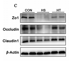
Journal: Journal Of Nutritional Biochemistry
アプリケーション: WB
交差性: Mouse
掲載日: 2025 Aug
-
Citation
-
Metabolites with Anti-Inflammatory Activities Isolated from the Mangrove Endophytic Fungus Dothiorella sp. ZJQQYZ-1
Author:
PMID: 40284726
Journal:
アプリケーション: WB
交差性: Mouse
掲載日: 2025 Apr
-
Citation
-
S-Adenosylmethionine Inhibits the Proliferation of Retinoblastoma Cell Y79, Induces Apoptosis and Cell Cycle Arrest of Y79 Cells by Inhibiting the Wnt2/β-Catenin Pathway
Author: Mushi Liu, Youchaou Mobet, Hong Shen
PMID: 39362212
Journal: Archivum Immunologiae Et Therapiae Experimentalis
アプリケーション: WB
交差性: Human
掲載日: 2024 Oct
-
Citation
-
Jintiange capsule ameliorates glucocorticoid-induced osteonecrosis of the femoral head in rats by regulating the activity and differentiation of BMSCs
Author: Xu Hui,et al
PMID: 39262662
Journal: Journal Of Traditional And Complementary Medicine
アプリケーション: WB
交差性: Rat
掲載日: 2024 Mar
-
Citation
-
BZW2 promotes malignant progression in lung adenocarcinoma through enhancing the ubiquitination and degradation of GSK3β
Author: Jin Kai,et al
PMID: 38424042
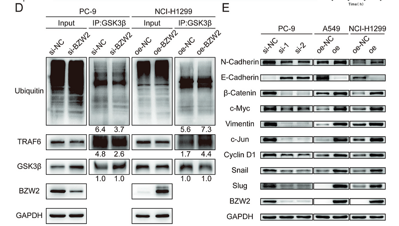
Journal: Cell Death Discovery
アプリケーション: WB
交差性: Human
掲載日: 2024 Feb
-
Citation
-
Effects of Zinc Combined with Metformin on Zinc Homeostasis, Blood-Epididymal Barrier, and Epididymal Absorption in Male Diabetic Mice
Author: Zhang Menghui,et al
PMID: 38589680
Journal: Biological Trace Element Research
アプリケーション: WB,IF
交差性: Mouse
掲載日: 2024 Apr
-
Citation
-
FZD7, Regulated by Non-CpG Methylation, Plays an Important Role in Immature Porcine Sertoli Cell Proliferation
Author: Yang, A., Yan, S., Yin, Y., Chen, C., Tang, X., Ran, M., & Chen, B.
PMID: 37047150
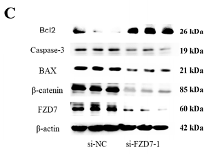
Journal: International Journal Of Molecular Sciences
アプリケーション: WB
交差性: Pig
掲載日: 2023 Mar
-
Citation
-
Tris (1, 3-dichloro-2-propyl) Phosphate Inhibits Early Embryonic Development by Binding to Gsk-3β Protein in Zebrafish
Author:
PMID: 37267805
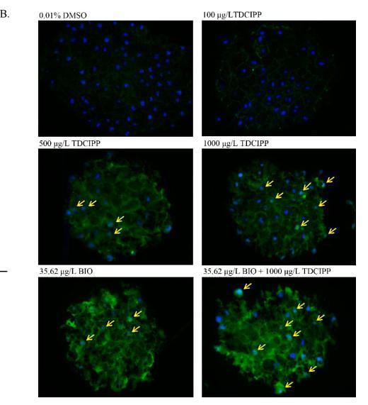
Journal: Aquatic Toxicology
アプリケーション: IF-cell
交差性: Zebrafish
掲載日: 2023 Jul
-
Citation
-
Transplantation of Menstrual Blood-Derived Mesenchymal Stem Cells Promotes the Repair of LPS-Induced Acute Lung Injury
Author: Charlie Xiang
PMID: 28346367
Journal: International Journal Of Molecular Sciences
アプリケーション: WB
交差性: Mouse
掲載日: 2017 Mar
-
Citation
-
Induction of VEGFA and Snail-1 by meningitic Escherichia coli mediates disruption of the blood-brain barrier
Author: Xiangru Wang
PMID: 27588479
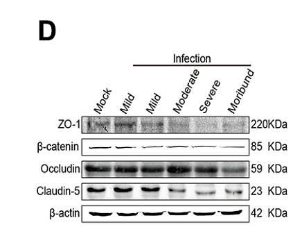
Journal: Oncotarget
アプリケーション: WB,IHC-P
交差性: Escherichia coli
掲載日: 2016 Sept
-
Citation
-
All-Trans Retinoic Acid-Induced Deficiency of the Wnt/β-Catenin Pathway Enhances Hepatic Carcinoma Stem Cell Differentiation
Author: Meizhang Li
PMID: 26571119
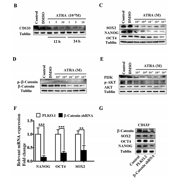
Journal: Public Library Of Science One
アプリケーション: WB
交差性: Human
掲載日: 2015 Nov
-
Citation
Alternative Products
同じターゲット & 同じ経路の製品
Beta Catenin Mouse Monoclonal Antibody [10-C0-B7]
Application: WB,IF-Cell,IHC-P,FC
Reactivity: Human,Mouse,Rat
Conjugate: unconjugated
Phospho-Beta Catenin (S552) Recombinant Rabbit Monoclonal Antibody [PSH08-72] - BSA and Azide free
Application: WB,IF-Cell,IHC-P,FC
Reactivity: Human,Mouse,Rat
Conjugate: unconjugated
Beta Catenin Mouse Monoclonal Antibody [A6-F8]
Application: WB,IF-Cell,IHC-P,FC,IF-Tissue,ChIP
Reactivity: Human,Mouse,Rat
Conjugate: unconjugated
Phospho-Beta Catenin (T41/S45) Recombinant Rabbit Monoclonal Antibody [JE54-02]
Application: WB,IHC-P
Reactivity: Human,Mouse,Rat
Conjugate: unconjugated
Phospho-Beta Catenin (S552) Recombinant Rabbit Monoclonal Antibody [PSH08-72]
Application: WB,IF-Cell,IHC-P,FC
Reactivity: Human,Mouse,Rat
Conjugate: unconjugated
Beta Catenin Recombinant Mouse Monoclonal Antibody [A6-F8-R] - BSA and Azide free
Application: WB,IF-Cell,FC
Reactivity: Human,Mouse,Rat
Conjugate: unconjugated
Beta Catenin Recombinant Rabbit Monoclonal Antibody [SA30-04] - BSA and Azide free
Application: WB,IHC-P,IF-Tissue,IP,IF-Cell,IHC-Fr,FC
Reactivity: Human,Mouse,Rat
Conjugate: unconjugated
Beta Catenin Recombinant Rabbit Monoclonal Antibody [SA30-04]
Application: WB,IHC-P,IF-Tissue,IP,mIHC,IF-Cell,IHC-Fr,FC
Reactivity: Human,Mouse,Rat
Conjugate: unconjugated
Beta Catenin Rabbit Polyclonal Antibody
Application: WB,IHC-P,FC,IF-Cell,IF-Tissue
Reactivity: Human,Mouse,Rat
Conjugate: unconjugated
Phospho-Beta Catenin (S33 + S37) Recombinant Rabbit Monoclonal Antibody [JE59-59]
Application: WB
Reactivity: Human,Rat,Mouse
Conjugate: unconjugated
iFluor™ 488 Conjugated Beta Catenin Recombinant Rabbit Monoclonal Antibody [SA30-04]
Application: IF-Tissue,IF-Cell
Reactivity: Human,Mouse
Conjugate: iFluor™ 488
Beta Catenin Recombinant Mouse Monoclonal Antibody [A6-F8-R]
Application: WB,IF-Cell,FC
Reactivity: Human,Mouse,Rat
Conjugate: unconjugated
Beta Catenin Recombinant Rabbit Monoclonal Antibody
Application: mIHC
Reactivity: Human
Conjugate: unconjugated


