HMGB1 Recombinant Rabbit Monoclonal Antibody [SA39-03]

製品仕様
今すぐ注文,【现货】
問い合わせをクリックCatalog# ET1601-2
HMGB1 Recombinant Rabbit Monoclonal Antibody [SA39-03]
-
WB
-
IF-Cell
-
IF-Tissue
-
IHC-P
-
FC
-
IHC-Fr
-
Human
-
Mouse
-
Rat
概要
製品名
HMGB1 Recombinant Rabbit Monoclonal Antibody [SA39-03]
抗体の種類
Recombinant Rabbit monoclonal Antibody
免疫原
Synthetic peptide within Human HMGB1 aa 151-200 / 215.
種属反応性
Human, Mouse, Rat
検証された応用例
WB, IF-Cell, IF-Tissue, IHC-P, FC, IHC-Fr
分子量
Predicted band size: 25 kDa
陽性対照
HeLa, HepG2 cell lysate, HeLa cell lysate, HCT 116 cell lysate, A549 cell lysate, Jurkat cell lysate, C2C12 cell lysate, C6 cell lysate, Mouse thymus tissue lysate, Rat spleen tissue lysate, MCF-7, human kidney tissue, mouse brain tissue, mouse hippocampus tissue, rat brain tissue, rat hippocampus tissue, C2C12, C6, HCT 116.
結合
unconjugated
クローン番号
SA39-03
RRID
製品の特徴
形態
Liquid
保存方法
Shipped at 4℃. Store at +4℃ short term (1-2 weeks). It is recommended to aliquot into single-use upon delivery. Store at -20℃ long term.
保存用バッファー
1*TBS (pH7.4), 0.05% BSA, 40% Glycerol. Preservative: 0.05% Sodium Azide.
アイソタイプ
IgG
精製方法
Protein A affinity purified.
応用希釈度
-
WB
-
1:20,000-1:50,000
-
IF-Cell
-
1:500
-
IF-Tissue
-
1:2,000
-
IHC-P
-
1:5,000-1:20,000
-
FC
-
1:1,000
-
IHC-Fr
-
1:500
論文における応用例
| WB | 確認する 12 以下の論文 |
| IF | 確認する 10 以下の論文 |
| IF-Cell | 確認する 1 以下の論文 |
| IHC-P | 確認する 1 以下の論文 |
論文における種属
| Mouse | 確認する 14 以下の論文 |
| Human | 確認する 7 以下の論文 |
| Rat | 確認する 2 以下の論文 |
| Grass carp | 確認する 1 以下の論文 |
| Carp | 確認する 1 以下の論文 |
ターゲット
機能
High mobility group box 1 protein, also known as high-mobility group protein 1 (HMG-1) and amphoterin, is a protein that in humans is encoded by the HMGB1 gene. HMG-1 belongs to the high mobility group and contains a HMG-box domain. Like the histones, HMGB1 is among the most important chromatin proteins. In the nucleus HMGB1 interacts with nucleosomes, transcription factors, and histones. This nuclear protein organizes the DNA and regulates transcription. After binding, HMGB1 bends DNA, which facilitates the binding of other proteins. HMGB1 supports transcription of many genes in interactions with many transcription factors. It also interacts with nucleosomes to loosen packed DNA and remodel the chromatin. Contact with core histones changes the structure of nucleosomes. The presence of HMGB1 in the nucleus depends on posttranslational modifications. When the protein is not acetylated, it stays in the nucleus, but hyperacetylation on lysine residues causes it to translocate into the cytosol. HMGB1 has been shown to play an important role in helping the RAG endonuclease form a paired complex during V(D)J recombination.
背景文献
1. "Novel role of PKR in inflammasome activation and HMGB1 release." Lu B., Nakamura T., Inouye K., Li J., Tang Y., Lundbaeck P., Valdes-Ferrer S.I., Olofsson P.S., Kalb T., Roth J., Zou Y., Erlandsson-Harris H., Yang H., Ting J.P., Wang H., Andersson U., Antoine D.J., Chavan S.S., Hotamisligil G.S., Tracey K.J. Nature 488:670-674(2012).
2. "The genetic variation of the human HMGB1 gene." Kornblit B., Munthe-Fog L., Petersen S., Madsen H., Vindeloev L., Garred P. Tissue Antigens 70:151-156(2007).
配列相同性
Belongs to the HMGB family.
組織特異性
Ubiquituous. Expressed in platelets.
翻訳後修飾
Phosphorylated at serine residues. Phosphorylation in both NLS regions is required for cytoplasmic translocation followed by secretion.; Acetylated on multiple sites upon stimulation with LPS. Acetylation on lysine residues in the nuclear localization signals (NLS 1 and NLS 2) leads to cytoplasmic localization and subsequent secretion (By similarity). Acetylation on Lys-3 results in preferential binding to DNA ends and impairs DNA bending activity (By similarity).; Reduction/oxidation of cysteine residues Cys-23, Cys-45 and Cys-106 and a possible intramolecular disulfide bond involving Cys-23 and Cys-45 give rise to different redox forms with specific functional activities in various cellular compartments: 1- fully reduced HMGB1 (HMGB1C23hC45hC106h), 2- disulfide HMGB1 (HMGB1C23-C45C106h) and 3- sulfonyl HMGB1 (HMGB1C23soC45soC106so).; Poly-ADP-ribosylated by PARP1 when secreted following stimulation with LPS (By similarity).; In vitro cleavage by CASP1 is liberating a HMG box 1-containing peptide which may mediate immunogenic activity; the peptide antagonizes apoptosis-induced immune tolerance. Can be proteolytically cleaved by a thrombin:thrombomodulin complex; reduces binding to heparin and proinflammatory activities (By similarity).
亜細胞局在
Cytoplasm, Nucleus, Cell membrane, Secreted, Chromosome
別名
Amphoterin antibody
Chromosomal protein, nonhistone, HMG1 antibody
DKFZp686A04236 antibody
High mobility group 1 antibody
High mobility group box 1 antibody
High mobility group protein 1 antibody
High mobility group protein B1 antibody
high-mobility group (nonhistone chromosomal) protein 1 antibody
HMG-1 antibody
HMG1 antibody
展開Amphoterin antibody
Chromosomal protein, nonhistone, HMG1 antibody
DKFZp686A04236 antibody
High mobility group 1 antibody
High mobility group box 1 antibody
High mobility group protein 1 antibody
High mobility group protein B1 antibody
high-mobility group (nonhistone chromosomal) protein 1 antibody
HMG-1 antibody
HMG1 antibody
HMG3 antibody
HMGB 1 antibody
HMGB1 antibody
HMGB1_HUMAN antibody
NONHISTONE CHROMOSOMAL PROTEIN HMG1 antibody
SBP 1 antibody
Sulfoglucuronyl carbohydrate binding protein antibody
折りたたむ画像
-

Western blot analysis of HMGB1 on different lysates with Rabbit anti-HMGB1 antibody (ET1601-2) at 1/50,000 dilution and competitor's antibody at 1/10,000 dilution.
Lane 1: HepG2 cell lysate
Lane 2: HeLa cell lysate
Lane 3: HCT 116 cell lysate
Lane 4: A549 cell lysate
Lane 5: Jurkat cell lysate
Lane 6: C2C12 cell lysate
Lane 7: C6 cell lysate
Lysates/proteins at 15 µg/Lane.
Predicted band size: 25 kDa
Observed band size: 25 kDa
Exposure time: 21 seconds; ECL: K1801;
4-20% SDS-PAGE gel.
Proteins were transferred to a PVDF membrane and blocked with 5% NFDM/TBST for 1 hour at room temperature. The primary antibody (ET1601-2) at 1/50,000 dilution and competitor's antibody at 1/10,000 dilution were used in 5% NFDM/TBST at 4℃ overnight. Goat Anti-Rabbit IgG - HRP Secondary Antibody (HA1001) at 1/50,000 dilution was used for 1 hour at room temperature. -
![<span style="font-weight: bold;">☑ Knockout (KO)</span><br /><br />Western blot analysis of HMGB1 with anti-HMGB1 antibody [SA39-03] (<a href="/products/ET1601-2" style="font-weight: bold;text-decoration: underline;">ET1601-2</a>) at 1/50,000 dilution.<br /><br />Lane 1: Wild-type Raw264.7 whole cell lysate.<br />Lane 2: HMGB1 knockout Raw264.7 whole cell lysate.<br /><br />Proteins were transferred to a PVDF membrane and blocked with 5% NFDM in TBST for 1 hour at room temperature. The primary Anti-HMGB1 antibody (<a href="/products/ET1601-2" style="font-weight: bold;text-decoration: underline;">ET1601-2</a>, 1/50,000) and Anti-HSP90 antibody (<a href="/products/ET1605-56" style="font-weight: bold;text-decoration: underline;">ET1605-56</a>, 1/10,000) were used in 5% BSA at room temperature for 2 hours. Goat Anti-Rabbit IgG H&L (HRP) Secondary Antibody (<a href="/products/HA1001" style="font-weight: bold;text-decoration: underline;">HA1001</a>) at 1/50,000 dilution was used for 1 hour at room temperature.<br /><br />Cell lysate was provided by Ubigene Biosciences (Ubigene Biosciences Co., Ltd., Guangzhou, China).](https://storage.huabio.cn/huabio/productImg/ET1601-2_2.jpg?v=20251127204004)
☑ Knockout (KO)
Western blot analysis of HMGB1 with anti-HMGB1 antibody [SA39-03] (ET1601-2) at 1/50,000 dilution.
Lane 1: Wild-type Raw264.7 whole cell lysate.
Lane 2: HMGB1 knockout Raw264.7 whole cell lysate.
Proteins were transferred to a PVDF membrane and blocked with 5% NFDM in TBST for 1 hour at room temperature. The primary Anti-HMGB1 antibody (ET1601-2, 1/50,000) and Anti-HSP90 antibody (ET1605-56, 1/10,000) were used in 5% BSA at room temperature for 2 hours. Goat Anti-Rabbit IgG H&L (HRP) Secondary Antibody (HA1001) at 1/50,000 dilution was used for 1 hour at room temperature.
Cell lysate was provided by Ubigene Biosciences (Ubigene Biosciences Co., Ltd., Guangzhou, China). -

Western blot analysis of HMGB1 on different lysates with Rabbit anti-HMGB1 antibody (ET1601-2) at 1/20,000 dilution.
Lane 1: HeLa cell lysate (20 µg/Lane)
Lane 2: HCT 116 cell lysate (20 µg/Lane)
Lane 3: A549 cell lysate (20 µg/Lane)
Lane 4: HepG2 cell lysate (20 µg/Lane)
Lane 5: Jurkat cell lysate (20 µg/Lane)
Lane 6: Mouse thymus tissue lysate (20 µg/Lane)
Lane 7: Rat spleen tissue lysate (30 µg/Lane)
Predicted band size: 25 kDa
Observed band size: 25 kDa
Exposure time: 3 minutes 10 seconds; ECL: K1801;
4-20% SDS-PAGE gel.
Proteins were transferred to a PVDF membrane and blocked with 5% NFDM/TBST for 1 hour at room temperature. The primary antibody (ET1601-2) at 1/20,000 dilution was used in 5% NFDM/TBST at 4℃ overnight. Goat Anti-Rabbit IgG - HRP Secondary Antibody (HA1001) at 1/50,000 dilution was used for 1 hour at room temperature. -

Immunocytochemistry analysis of HeLa cells labeling HMGB1 with Rabbit anti-HMGB1 antibody (ET1601-2) at 1/500 dilution and competitor's antibody at 1/250 dilution.
Cells were fixed in 4% paraformaldehyde for 20 minutes at room temperature, permeabilized with 0.1% Triton X-100 in PBS for 5 minutes at room temperature, then blocked with 1% BSA in 10% negative goat serum for 1 hour at room temperature. Cells were then incubated with Rabbit anti-HMGB1 antibody (ET1601-2) at 1/500 dilution and competitor's antibody at 1/250 dilution in 1% BSA in PBST overnight at 4 ℃. Goat Anti-Rabbit IgG H&L (iFluor™ 488, HA1121) was used as the secondary antibody at 1/1,000 dilution. PBS instead of the primary antibody was used as the secondary antibody only control. Nuclear DNA was labelled in blue with DAPI.
Beta tubulin (M1305-2, red) was stained at 1/100 dilution overnight at +4℃. Goat Anti-Mouse IgG H&L (iFluor™ 594, HA1126) was used as the secondary antibody at 1/1,000 dilution. -

Immunocytochemistry analysis of MCF-7 cells labeling HMGB1 with Rabbit anti-HMGB1 antibody (ET1601-2) at 1/100 dilution.
Cells were fixed in 4% paraformaldehyde for 10 minutes at 37 ℃, permeabilized with 0.05% Triton X-100 in PBS for 20 minutes, and then blocked with 2% negative goat serum for 30 minutes at room temperature. Cells were then incubated with Rabbit anti-HMGB1 antibody (ET1601-2) at 1/100 dilution in 2% negative goat serum overnight at 4 ℃. Goat Anti-Rabbit IgG H&L (iFluor™ 488, HA1121) was used as the secondary antibody at 1/1,000 dilution. PBS instead of the primary antibody was used as the secondary antibody only control. Nuclear DNA was labelled in blue with DAPI. -

Immunohistochemical analysis of paraffin-embedded human kidney tissue with Rabbit anti-HMGB1 antibody (ET1601-2) at 1/20,000 dilution.
The section was pre-treated using heat mediated antigen retrieval with sodium citrate buffer (pH 6.0) (high pressure) for 2 minutes. The tissues were blocked in 1% BSA for 20 minutes at room temperature, washed with ddH2O and PBS, and then probed with the primary antibody (ET1601-2) at 1/20,000 dilution for 1 hour at room temperature. The detection was performed using an HRP conjugated compact polymer system. DAB was used as the chromogen. Tissues were counterstained with hematoxylin and mounted with DPX. -

Immunohistochemical analysis of paraffin-embedded mouse brain tissue with Rabbit anti-HMGB1 antibody (ET1601-2) at 1/20,000 dilution.
The section was pre-treated using heat mediated antigen retrieval with sodium citrate buffer (pH 6.0) (high pressure) for 2 minutes. The tissues were blocked in 1% BSA for 20 minutes at room temperature, washed with ddH2O and PBS, and then probed with the primary antibody (ET1601-2) at 1/20,000 dilution for 1 hour at room temperature. The detection was performed using an HRP conjugated compact polymer system. DAB was used as the chromogen. Tissues were counterstained with hematoxylin and mounted with DPX. -

Immunohistochemical analysis of paraffin-embedded mouse hippocampus tissue with Rabbit anti-HMGB1 antibody (ET1601-2) at 1/20,000 dilution.
The section was pre-treated using heat mediated antigen retrieval with sodium citrate buffer (pH 6.0) (high pressure) for 2 minutes. The tissues were blocked in 1% BSA for 20 minutes at room temperature, washed with ddH2O and PBS, and then probed with the primary antibody (ET1601-2) at 1/20,000 dilution for 1 hour at room temperature. The detection was performed using an HRP conjugated compact polymer system. DAB was used as the chromogen. Tissues were counterstained with hematoxylin and mounted with DPX. -

Immunohistochemical analysis of paraffin-embedded rat brain tissue with Rabbit anti-HMGB1 antibody (ET1601-2) at 1/20,000 dilution.
The section was pre-treated using heat mediated antigen retrieval with sodium citrate buffer (pH 6.0) (high pressure) for 2 minutes. The tissues were blocked in 1% BSA for 20 minutes at room temperature, washed with ddH2O and PBS, and then probed with the primary antibody (ET1601-2) at 1/20,000 dilution for 1 hour at room temperature. The detection was performed using an HRP conjugated compact polymer system. DAB was used as the chromogen. Tissues were counterstained with hematoxylin and mounted with DPX. -

Immunohistochemical analysis of paraffin-embedded rat hippocampus tissue with Rabbit anti-HMGB1 antibody (ET1601-2) at 1/20,000 dilution.
The section was pre-treated using heat mediated antigen retrieval with sodium citrate buffer (pH 6.0) (high pressure) for 2 minutes. The tissues were blocked in 1% BSA for 20 minutes at room temperature, washed with ddH2O and PBS, and then probed with the primary antibody (ET1601-2) at 1/20,000 dilution for 1 hour at room temperature. The detection was performed using an HRP conjugated compact polymer system. DAB was used as the chromogen. Tissues were counterstained with hematoxylin and mounted with DPX. -

Immunocytochemistry analysis of C2C12 cells labeling HMGB1 with Rabbit anti-HMGB1 antibody (ET1601-2) at 1/500 dilution.
Cells were fixed in 4% paraformaldehyde for 20 minutes at room temperature, permeabilized with 0.1% Triton X-100 in PBS for 5 minutes at room temperature, then blocked with 1% BSA in 10% negative goat serum for 1 hour at room temperature. Cells were then incubated with Rabbit anti-HMGB1 antibody (ET1601-2) at 1/500 dilution in 1% BSA in PBST overnight at 4 ℃. Goat Anti-Rabbit IgG H&L (iFluor™ 488, HA1121) was used as the secondary antibody at 1/1,000 dilution. PBS instead of the primary antibody was used as the secondary antibody only control. Nuclear DNA was labelled in blue with DAPI.
Beta tubulin (M1305-2, red) was stained at 1/100 dilution overnight at +4℃. Goat Anti-Mouse IgG H&L (iFluor™ 594, HA1126) was used as the secondary antibody at 1/1,000 dilution. -

Immunocytochemistry analysis of C6 cells labeling HMGB1 with Rabbit anti-HMGB1 antibody (ET1601-2) at 1/500 dilution.
Cells were fixed in 4% paraformaldehyde for 20 minutes at room temperature, permeabilized with 0.1% Triton X-100 in PBS for 5 minutes at room temperature, then blocked with 1% BSA in 10% negative goat serum for 1 hour at room temperature. Cells were then incubated with Rabbit anti-HMGB1 antibody (ET1601-2) at 1/500 dilution in 1% BSA in PBST overnight at 4 ℃. Goat Anti-Rabbit IgG H&L (iFluor™ 488, HA1121) was used as the secondary antibody at 1/1,000 dilution. PBS instead of the primary antibody was used as the secondary antibody only control. Nuclear DNA was labelled in blue with DAPI.
Beta tubulin (M1305-2, red) was stained at 1/100 dilution overnight at +4℃. Goat Anti-Mouse IgG H&L (iFluor™ 594, HA1126) was used as the secondary antibody at 1/1,000 dilution. -

Flow cytometric analysis of HCT 116 cells labeling HMGB1.
Cells were fixed and permeabilized. Then stained with the primary antibody (ET1601-2, 1/1,000) (red) compared with Rabbit IgG Isotype Control (green). After incubation of the primary antibody at +4℃ for an hour, the cells were stained with a iFluor™ 488 conjugate-Goat anti-Rabbit IgG Secondary antibody (HA1121) at 1/1,000 dilution for 30 minutes at +4℃. Unlabelled sample was used as a control (cells without incubation with primary antibody; black). -

Application: IF-Tissue
Species: Mouse
Site: brain
Sample: Paraffin-embedded section
Antibody concentration: 1/2,000 -

Application: IHC-Fr
Species: Mouse
Site: brain
Sample: Frozen section
Antibody concentration: 1/500
Antigen retrieval: Not required -

Western blot analysis of HMGB1 on Mouse HMGB1 recombinant protein with Rabbit anti-HMGB1 antibody (ET1601-2) at 1/1,000 dilution.
Lysates/proteins at 50 ng/Lane.
Exposure time: 59 seconds; ECL: K1801;
4-20% SDS-PAGE gel.
Proteins were transferred to a PVDF membrane and blocked with 5% NFDM/TBST for 1 hour at room temperature. The primary antibody (ET1601-2) at 1/1,000 dilution was used in primary antibody dilution (K1803) at 4℃ overnight. Goat Anti-Rabbit IgG - HRP Secondary Antibody (HA1001) at 1/50,000 dilution was used for 1 hour at room temperature.
ご注意ください: All products are "FOR RESEARCH USE ONLY AND ARE NOT INTENDED FOR DIAGNOSTIC OR THERAPEUTIC USE"
論文での実績
-
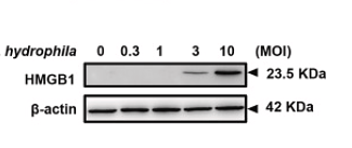
Teleost HMGB1 paralogues mediate protective autophagy by interacting with NOD2 and ATG16L1 and activating ROS/Akt/mTOR pathway against Aeromonas hydrophila infection
Author: Hong Zhou
PMID: 41015164
Journal: Fish & Shellfish Immunology
アプリケーション: WB
交差性: Carp
掲載日: 2025 Sept
-
Citation
-
Platform-based BST-2-targeted microbubbles enhance HIFU therapy and effectively inhibit prostate cancer residual growth
Author: Hang Zhou, Jia Tang, Yujie Wan, Jiaqi Gong, Qizhi Wang, Kaifeng Huang, Ruixia Hong, Xinzhi Xu, Fang Li
PMID: 40436774
Journal: International Journal of Hyperthermia
アプリケーション: IF
交差性: Mouse
掲載日: 2025 May
-
Citation
-
FKBP5 Mediates Alveolar Fibroblast Necroptosis During Acute Respiratory Distress Syndrome
Author: Dong Zhang, Wei Liu, Ting Sun, Yangyang Xiao, Qiuwen Chen, Xiao Huang, Xiaozhi Wang, Qian Qi, Hao Wang, Tao Wang
PMID: 40525648
Journal: Cell Proliferation
アプリケーション: WB
交差性: Mouse,Human
掲載日: 2025 Jun
-
Citation
-
Spatiotemporal-Controlled Nanoagonists Triggered STING Activation for Cascade-Amplified Photothermal Metalloimmunotherapy
Author: Min Mu, Hui Li, Susu Xiao, Chenqian Feng, Bo Chen, Rangrang Fan, Nianyong Chen, Gang Guo
PMID: 40629961
Journal: Biomacromolecules
アプリケーション: IF
交差性: Mouse
掲載日: 2025 Jul
-
Citation
-
Environmental enrichment attenuates social isolation-exacerbated postoperative cognitive dysfunction in aged mice via inhibition of RAGE-HMGB1 proinflammatory signaling.
Author: Sha Li, Hongyan Wang, Mingzhe Qin, Wei Huang, Huifang Gao, Xiaoyang Song, Xiaolong Chen, Bixi Li
PMID: 40653061
Journal: Brain Research Bulletin
アプリケーション: WB
交差性: Mouse
掲載日: 2025 Jul
-
Citation
-
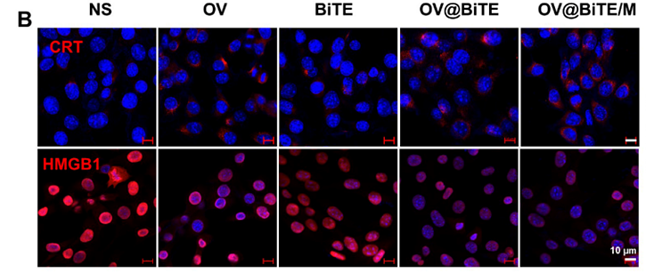
A fusion of oncolytic viruses and bispecific T-cell engagers for triple negative breast cancer immunotherapy
Author: Min Mu, Hui Li, Guoqing Wang, Bo Chen, Rangrang Fan, Nianyong Chen, Bingwen Zou, Aiping Tong, Gang Guo
PMID: 25NOPMID25032701
Journal: Chemical Engineering Journal
アプリケーション: IF
交差性: Mouse
掲載日: 2025 Feb
-
Citation
-
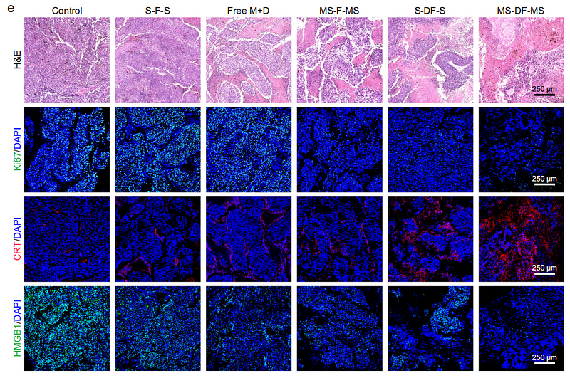
Cascaded immunotherapy with implantable dual-drug depots sequentially releasing STING agonists and apoptosis inducers
Author: Li Kai, Yu Xuan, Xu Yanteng, Wang Haixia, Liu Zheng, Wu Chong, Luo Xing, Xu Jiancheng, Fang Youqiang, Ju Enguo, Lv Shixian, Chan Hon Fai, Lao Yeh-Hsing, He Weiling, Tao Yu, Li Mingqiang
PMID: 39952937
Journal: Nature Communications
アプリケーション: IF
交差性: Mouse
掲載日: 2025 Feb
-
Citation
-
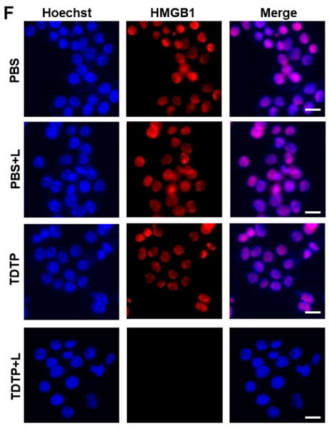
Harnessing Membrane-Targeting Photosensitizers to Evoke Ferroptosis and Pyroptosis Dual-Modal Cell Death for Antitumor Therapy
Author: Ying Yin, Xiang Cheng, Duoyang Fan, Yanpeng Fang, Haohan Li, Xingru Zhou, Hongqi Guo, Wenbin Zeng, Fei Chen
PMID: 40875307
Journal: Molecular Pharmaceutics
アプリケーション: IF
交差性: Mouse
掲載日: 2025 Aug
-
Citation
-
Stromal Reprogramming Optimizes KRAS-Specific Chemotherapy Inducing Antitumor Immunity in Pancreatic Cancer
Author: Qinglian Hu, Jiayu Feng, Lulu Qi, Yuanxiang Jin
PMID: 39480275
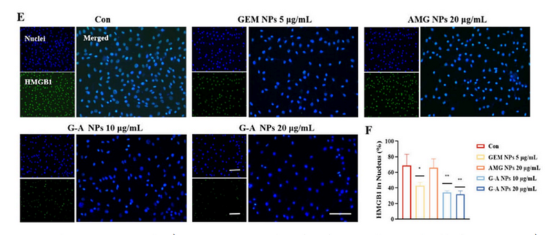
Journal: ACS Applied Materials & Interfaces
アプリケーション:
交差性:
掲載日: 2024 Oct
-
Citation
-
Triggering Pyroptosis by Doxorubicin-Loaded Multifunctional Nanoparticles in Combination with Decitabine for Breast Cancer Chemoimmunotherapy
Author: Xueyan Hou,et al
PMID: 39413405
Journal: Biological And Medical Applications Of Materials And Interfaces
アプリケーション: IF
交差性: Human
掲載日: 2024 Oct
-
Citation
-
Ursolic acid improves necroptosis via STAT3 signaling in intestinal ischemia/reperfusion injury
Author: Shi Yajing,et al
PMID: 38971110
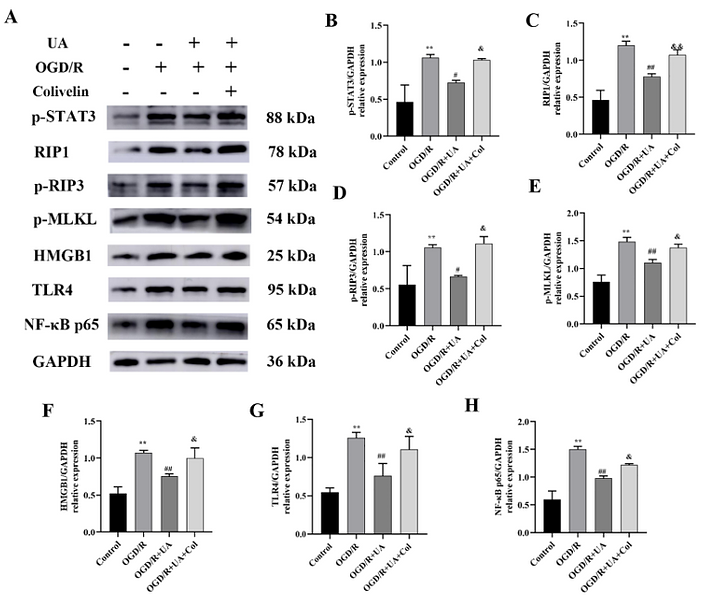
Journal: International Immunopharmacology
アプリケーション: WB
交差性: Mouse
掲載日: 2024 Jul
-
Citation
-
A Self-Cascade Oxygen-Generating Nanomedicine for Multimodal Tumor Therapy
Author: Jingyuan Zhao, Qi Sun, Dongze Mo, Jiayuan Feng, Yuting Wang, Tong Li, Yihong Zhang, Hui Wei
PMID: 38966876
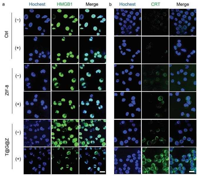
Journal: Small
アプリケーション:
交差性:
掲載日: 2024 Jul
-
Citation
-
Injectable alginate hydrogel promotes antitumor immunity through glucose oxidase and Fe3+ amplified RSL3-induced ferroptosis
Author: Chen K, Gu L, Zhang Q, et al
PMID: 38142082
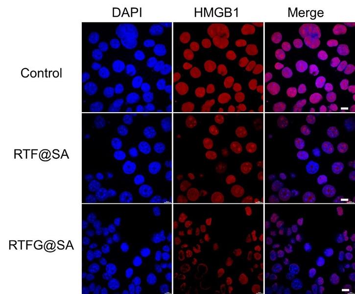
Journal: Carbohydrate Polymers
アプリケーション: IF-Cell
交差性: Mouse
掲載日: 2024 Feb
-
Citation
-
A sodium alginate-based injectable hydrogel system for locoregional treatment of colorectal cancer by eliciting pyroptosis and apoptosis
Author: Yongli Shi, Jingya Zhao, Suyue Xu, Huiqing Zhu, Yuxin Wang, Bingqian Zhao, Zeyu Sun, Sisi He, Xueyan Hou
PMID: 39746425
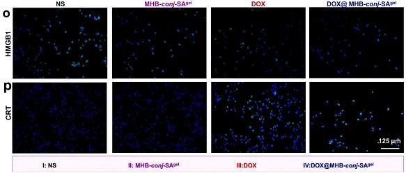
Journal: International Journal Of Biological Macromolecules
アプリケーション: WB
交差性: Mouse
掲載日: 2024 Dec
-
Citation
-
EZH2/miR-142-3p/HMGB1 axis mediates chondrocyte pyroptosis by regulating endoplasmic reticulum stress in knee osteoarthritis
Author: Haibo Si,et al
PMID: 39704001
Journal: Chinese Medical Journal
アプリケーション: WB
交差性: Human
掲載日: 2024 Dec
-
Citation
-
Fingolimod Alleviates Inflammation after Cerebral Ischemia via HMGB1/TLR4/NF‑κB Signaling Pathway
Author: Xing Yao,et al
PMID: 39207074
Journal: Journal Of Integrative Neuroscience
アプリケーション: IHC-P,WB
交差性: Rat
掲載日: 2024 Aug
-
Citation
-
Targeting the MCP-GPX4/HMGB1 Axis for Effectively Triggering Immunogenic Ferroptosis in Pancreatic Ductal Adenocarcinoma
Author: Ge Li, Chengyu Liao, Jiangzhi Chen, Zuwei Wang, Shuncang Zhu, Jianlin Lai, Qiaowei Li, Yinhao Chen, Dihan Wu, Jianbo Li, Yi Huang, Yifeng Tian, Yanling Chen, Shi Chen
PMID: 38593415
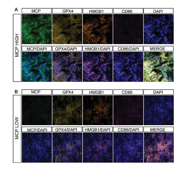
Journal: Advanced Science
アプリケーション: IF
交差性: Human
掲載日: 2024 Apr
-
Citation
-
Lipid–Polymer Hybrid Nanoparticles with Both PD-L1 Knockdown and Mild Photothermal Effect for Tumor Photothermal Immunotherapy
Author:
PMID: 37605506
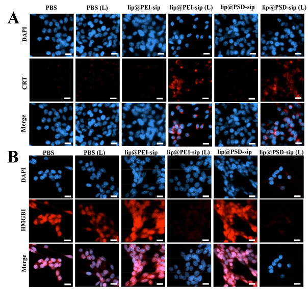
Journal: ACS Applied Materials & Interfaces
アプリケーション: IF
交差性: Mouse
掲載日: 2023 Sept
-
Citation
-
Grass carp Il-2 promotes neutrophil extracellular traps formation via inducing ROS production and autophagy in vitro
Author: Lv M, Wang Y, Yu J, et al
PMID: 38040137
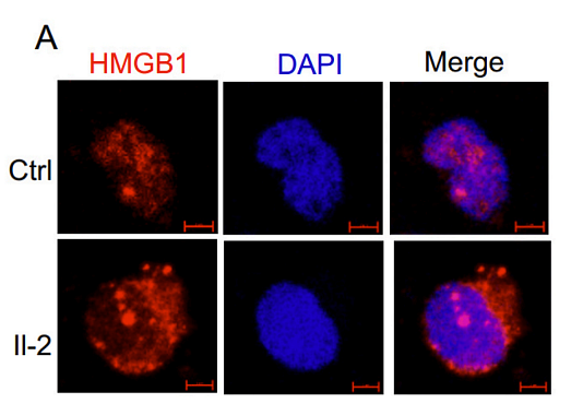
Journal: Fish & Shellfish Immunology
アプリケーション: IF
交差性: Grass carp
掲載日: 2023 Nov
-
Citation
-
mTOR signaling pathway regulates embryonic development and rapid growth of triploid crucian carp
Author: Huang Z , Dai L, Peng F,et al
PMID: NO PMID202311
Journal: Aquaculture Reports
アプリケーション:
交差性: Mouse
掲載日: 2023 Nov
-
Citation
-
Reduced HMGB1 expression contributed to lapatinib-induced cutaneous injury
Author:
PMID: NA1
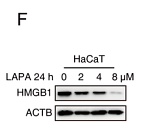
Journal: Research Square
アプリケーション: WB
交差性: Human
掲載日: 2022 Mar
-
Citation
-
SARS-CoV-2 ORF3a induces RETREG1/FAM134B dependent reticulophagy and triggers sequential ER stress and inflammatory responses during SARS-CoV-2 infection
Author: Zhang, X., Yang, Z., Pan, T., Long, X., Sun, Q., Wang, P. H., Li, X., & Kuang, E.
PMID: 35239449
Journal: Autophagy
アプリケーション: WB
交差性: Human
掲載日: 2022 Mar
-
Citation
-
Ginsenoside Rh2 Inhibits NLRP3 Inflammasome Activation and Improves Exosomes to Alleviate Hypoxia-Induced Myocardial Injury
Author:
PMID: 35865525
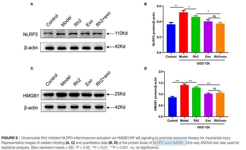
Journal: Frontiers In Immunology
アプリケーション: WB
交差性: Rat
掲載日: 2022 Jul
-
Citation
-
Decreased HMGB1 expression contributed to cutaneous toxicity caused by lapatinib
Author:
PMID: 35617997
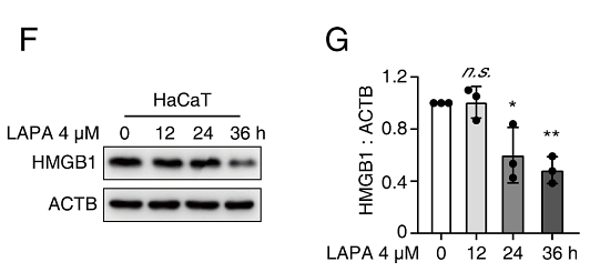
Journal: Biochemical Pharmacology
アプリケーション: WB
交差性: Human
掲載日: 2022 Jul
-
Citation
-
Autophagic degradation of CCN2 (cellular communication network factor 2) causes cardiotoxicity of sunitinib
Author: Xu, Z., Jin, Y., Gao, Z., Zeng, Y., Du, J., Yan, H., Chen, X., Ping, L., Lin, N., Yang, B., He, Q., & Luo, P.
PMID: 34432562
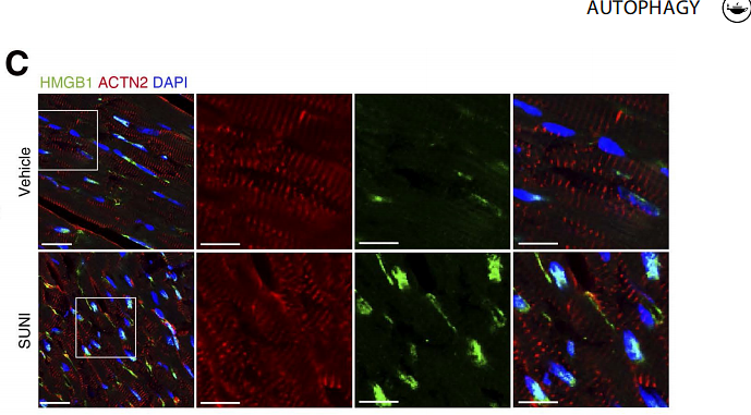
Journal: Autophagy
アプリケーション: IF
交差性: Mouse
掲載日: 2021 Aug
-
Citation
-
Mesenchymal Stem Cells Attenuate Diabetic Lung Fibrosis via Adjusting Sirt3-Mediated Stress Responses in Rats
Author: Yang Chen, Fuping Zhang, Di Wang, Lan Li, Haibo Si, Chengshi Wang, Jingping Liu, Younan Chen, Jingqiu Cheng, Yanrong Lu
PMID: 32089781
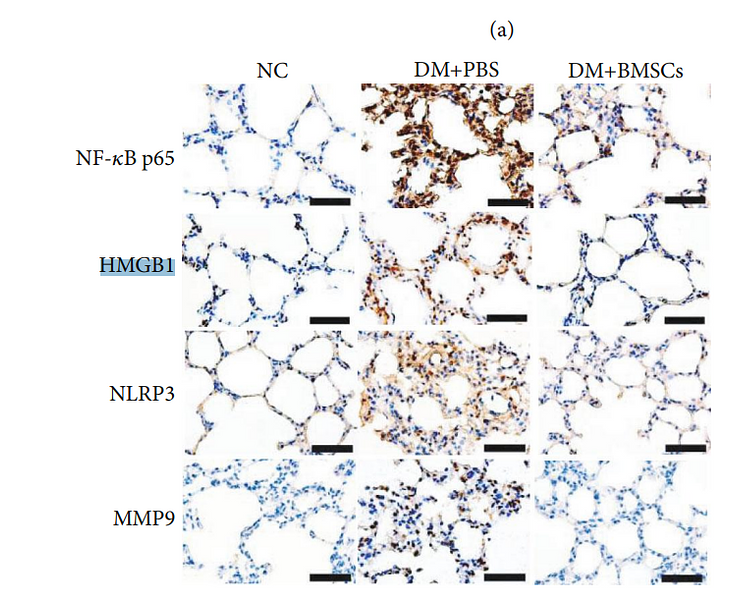
Journal: Oxidative Medicine And Cellular Longevity
アプリケーション: WB
交差性: Mouse
掲載日: 2020 Feb
-
Citation
同じターゲット & 同じ経路の製品
HMGB1 Rabbit Polyclonal Antibody
Application: WB,IF-Cell,IHC-P,FC
Reactivity: Human,Mouse,Rat
Conjugate: unconjugated
HMGB1 Recombinant Rabbit Monoclonal Antibody [SA39-03] - BSA and Azide free
Application: WB,IF-Cell,IF-Tissue,IHC-P,FC,IHC-Fr
Reactivity: Human,Mouse,Rat
Conjugate: unconjugated
HMGB1 Rabbit Polyclonal Antibody
Application: WB,IF-Cell,IHC-P,FC
Reactivity: Human,Mouse,Rat
Conjugate: unconjugated


