HER2 / ErbB2 Rabbit Polyclonal Antibody
製品仕様
今すぐ注文,【现货】
問い合わせをクリックCatalog# ER0106
HER2 / ErbB2 Rabbit Polyclonal Antibody
-
WB
-
IHC-P
-
Human
-
unconjugated
概要
製品名
HER2 / ErbB2 Rabbit Polyclonal Antibody
抗体の種類
Rabbit Polyclonal Antibody
免疫原
Synthetic peptide within Human HER2 aa 1-50 / 1,255.
種属反応性
Human
検証された応用例
WB, IHC-P
分子量
Predicted band size: 138 kDa
陽性対照
SK-Br-3 cell lysate, SKOV-3 cell lysate, 4T1 cell lysate, human breast cancer tissue, human stomach carcinoma tissue, human placenta tissue.
結合
unconjugated
RRID
製品の特徴
形態
Liquid
保存方法
Shipped at 4℃. Store at +4℃ short term (1-2 weeks). It is recommended to aliquot into single-use upon delivery. Store at -20℃ long term.
保存用バッファー
1*PBS (pH7.4), 0.2% BSA, 40% Glycerol. Preservative: 0.05% Sodium Azide.
アイソタイプ
IgG
精製方法
Immunogen affinity purified.
応用希釈度
-
WB
-
1:500-1:1,000
-
IHC-P
-
1:50-1:1,000
論文における応用例
| IHC-P | 確認する 1 以下の論文 |
| WB | 確認する 1 以下の論文 |
論文における種属
| Human | 確認する 2 以下の論文 |
ターゲット
機能
HER2 is a member of the human epidermal growth factor receptor (HER/EGFR/ERBB) family. Amplification or overexpression of this oncogene has been shown to play an important role in the development and progression of certain aggressive types of breast cancer. In recent years the protein has become an important biomarker and target of therapy for approx. 30% of breast cancer patients. Over-expression is also known to occur in ovarian, stomach, and aggressive forms of uterine cancer, such as uterine serous endometrial carcinoma. The expression of HER2 is regulated by signaling through estrogen receptors. Normally, estradiol and tamoxifen acting through the estrogen receptor down-regulate the expression of HER2. However, when the ratio of the coactivator AIB-3 exceeds that of the corepressor PAX2, the expression of HER2 is upregulated in the presence of tamoxifen, leading to tamoxifen-resistant breast cancer.
背景文献
1. "Endosomal transport of ErbB-2: mechanism for nuclear entry of the cell surface receptor." Giri D.K., Ali-Seyed M., Li L.Y., Lee D.F., Ling P., Bartholomeusz G., Wang S.C., Hung M.C. Mol. Cell. Biol. 25:11005-11018(2005)
2. "Brk is coamplified with ErbB2 to promote proliferation in breast cancer." Xiang B., Chatti K., Qiu H., Lakshmi B., Krasnitz A., Hicks J., Yu M., Miller W.T., Muthuswamy S.K.Proc. Natl. Acad. Sci. U.S.A. 105:12463-12468(2008)
3. "ErbB2-mediated Src and signal transducer and activator of transcription 3 activation leads to transcriptional up-regulation of p21Cip1 and chemoresistance in breast cancer cells." Hawthorne V.S., Huang W.C., Neal C.L., Tseng L.M., Hung M.C., Yu D. Mol. Cancer Res. 7:592-600(2009)
配列相同性
Belongs to the protein kinase superfamily. Tyr protein kinase family. EGF receptor subfamily.
組織特異性
Expressed in a variety of tumor tissues including primary breast tumors and tumors from small bowel, esophagus, kidney and mouth.
翻訳後修飾
Autophosphorylated. Autophosphorylation occurs in trans, i.e. one subunit of the dimeric receptor phosphorylates tyrosine residues on the other subunit (Probable). Ligand-binding increases phosphorylation on tyrosine residues. Signaling via SEMA4C promotes phosphorylation at Tyr-1248. Dephosphorylated by PTPN12.
亜細胞局在
Cell membrane.
UNIPROT #
別名
Verb b2 erythroblastic leukemia viral oncogene homolog 2, neuro/glioblastoma derived oncogene homolog antibody
C erb B2/neu protein antibody
CD340 antibody
CD340 antigen antibody
Cerb B2/neu protein antibody
CerbB2 antibody
Erb b2 receptor tyrosine kinase 2 antibody
ERBB2 antibody
ERBB2_HUMAN antibody
HER 2 antibody
展開Verb b2 erythroblastic leukemia viral oncogene homolog 2, neuro/glioblastoma derived oncogene homolog antibody
C erb B2/neu protein antibody
CD340 antibody
CD340 antigen antibody
Cerb B2/neu protein antibody
CerbB2 antibody
Erb b2 receptor tyrosine kinase 2 antibody
ERBB2 antibody
ERBB2_HUMAN antibody
HER 2 antibody
HER 2/NEU antibody
HER2 antibody
Herstatin antibody
Human epidermal growth factor receptor 2 antibody
Metastatic lymph node gene 19 protein antibody
MLN 19 antibody
MLN19 antibody
NEU antibody
NEU proto oncogene antibody
Neuro/glioblastoma derived oncogene homolog antibody
Neuroblastoma/glioblastoma derived oncogene homolog antibody
NGL antibody
p185erbB2 antibody
Proto-oncogene c-ErbB-2 antibody
Proto-oncogene Neu antibody
Receptor tyrosine-protein kinase erbB-2 antibody
TKR1 antibody
Tyrosine kinase type cell surface receptor HER2 antibody
Tyrosine kinase-type cell surface receptor HER2 antibody
V erb b2 avian erythroblastic leukemia viral oncogene homolog 2 (neuro/glioblastoma derived oncogene homolog) antibody
V erb b2 avian erythroblastic leukemia viral oncogene homolog 2 antibody
V erb b2 avian erythroblastic leukemia viral oncoprotein 2 antibody
V erb b2 erythroblastic leukemia viral oncogene homolog 2, neuro/glioblastoma derived oncogene homolog (avian) antibody
V erb b2 erythroblastic leukemia viral oncogene homolog 2, neuro/glioblastoma derived oncogene homolog antibody
Verb b2 erythroblastic leukemia viral oncogene homolog 2, neuro/glioblastoma derived oncogene homolog (avian) antibody
折りたたむ画像
-

Western blot analysis of HER2 / ErbB2 on different lysates with Rabbit anti-HER2 / ErbB2 antibody (ER0106) at 1/1,000 dilution.
Lane 1: SK-Br-3 cell lysate
Lane 2: SKOV-3 cell lysate
Lysates/proteins at 10 µg/Lane.
Predicted band size: 138 kDa
Observed band size: 180 kDa
Exposure time: 2 minutes;
6% SDS-PAGE gel.
Proteins were transferred to a PVDF membrane and blocked with 5% NFDM/TBST for 1 hour at room temperature. The primary antibody (ER0106) at 1/1,000 dilution was used in 5% NFDM/TBST at room temperature for 2 hours. Goat Anti-Rabbit IgG - HRP Secondary Antibody (HA1001) at 1:300,000 dilution was used for 1 hour at room temperature. -

Western blot analysis of HER2 / ErbB2 on 4T1 cell lysates with Rabbit anti-HER2 / ErbB2 antibody (ER0106) at 1/1,000 dilution.
Lysates/proteins at 20 µg/Lane.
Predicted band size: 138 kDa
Observed band size: 250 kDa
Exposure time: 25 seconds; ECL: K1801;
4-20% SDS-PAGE gel.
Proteins were transferred to a PVDF membrane and blocked with 5% NFDM/TBST for 1 hour at room temperature. The primary antibody (ER0106) at 1/1,000 dilution was used in 5% NFDM/TBST at 4℃ overnight. Goat Anti-Rabbit IgG - HRP Secondary Antibody (HA1001) at 1/50,000 dilution was used for 1 hour at room temperature. -

Immunohistochemical analysis of paraffin-embedded human breast cancer tissue with Rabbit anti-HER2 / ErbB2 antibody (ER0106) at 1/1,000 dilution.
The section was pre-treated using heat mediated antigen retrieval with Tris-EDTA buffer (pH 9.0) for 20 minutes. The tissues were blocked in 1% BSA for 20 minutes at room temperature, washed with ddH2O and PBS, and then probed with the primary antibody (ER0106) at 1/1,000 dilution for 1 hour at room temperature. The detection was performed using an HRP conjugated compact polymer system. DAB was used as the chromogen. Tissues were counterstained with hematoxylin and mounted with DPX. -

Immunohistochemical analysis of paraffin-embedded human stomach carcinoma tissue using anti-HER2 / ErbB2 antibody. The section was pre-treated using heat mediated antigen retrieval with Tris-EDTA buffer (pH 8.0-8.4) for 20 minutes.The tissues were blocked in 5% BSA for 30 minutes at room temperature, washed with ddH2O and PBS, and then probed with the primary antibody (ER0106, 1/50) for 30 minutes at room temperature. The detection was performed using an HRP conjugated compact polymer system. DAB was used as the chromogen. Tissues were counterstained with hematoxylin and mounted with DPX.
-

Immunohistochemical analysis of paraffin-embedded human placenta tissue using anti-HER2 / ErbB2 antibody. The section was pre-treated using heat mediated antigen retrieval with Tris-EDTA buffer (pH 8.0-8.4) for 20 minutes.The tissues were blocked in 5% BSA for 30 minutes at room temperature, washed with ddH2O and PBS, and then probed with the primary antibody (ER0106, 1/1,000) for 30 minutes at room temperature. The detection was performed using an HRP conjugated compact polymer system. DAB was used as the chromogen. Tissues were counterstained with hematoxylin and mounted with DPX.
ご注意ください: All products are "FOR RESEARCH USE ONLY AND ARE NOT INTENDED FOR DIAGNOSTIC OR THERAPEUTIC USE"
論文での実績
-
Assembly of Dye Molecules in Covalent Organic Frameworks for Enhanced Colorimetric Biosensing
Author: Lin Wang,et al
PMID: 39283703
Journal: Analytical Chemistry
アプリケーション:
交差性:
掲載日: 2024 Sep
-
Citation
-
Overexpression of miR-489-3p inhibits Proliferation and Migration of Non-Small Cell Lung Cancer cells by Suppressing the HER2/PI3K/AKT/Snail signaling Pathway
Author: Cheng Di,et al
PMID: 39224367
Journal: Heliyon
アプリケーション: WB
交差性: Human
掲載日: 2024 Aug
-
Citation
-
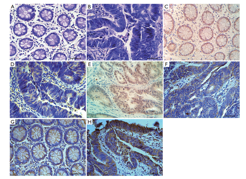
Expression, clinical significance and correlation of RUNX3 and HER2 in colorectal cancer
Author: Wu, Y., Xue, J., Li, Y., Wu, X., Qu, M., Xu, D., & Shi, Y.
PMID: 34532112

Journal: Journal Of Gastrointestinal Oncology
アプリケーション: IHC-P
交差性: Human
掲載日: 2021 Aug
-
Citation
Alternative Products
同じターゲット & 同じ経路の製品
HER2 / ErbB2 Recombinant Rabbit Monoclonal Antibody [PD00-53]
Application: IHC-P,WB,IF-Cell,FC,mIHC
Reactivity: Human
Conjugate: unconjugated
Phospho-HER2 / ErbB2 (Y1221 + Y1222) Recombinant Rabbit Monoclonal Antibody [JE44-12]
Application: WB
Reactivity: Human
Conjugate: unconjugated
Phospho-HER2 / ErbB2 (Y1248) Recombinant Rabbit Monoclonal Antibody [PSH04-03]
Application: WB,IF-Cell
Reactivity: Human
Conjugate: unconjugated
Phospho-HER2 / ErbB2 (Y1139) Recombinant Rabbit Monoclonal Antibody [JE60-90]
Application: WB,IF-Cell,FC
Reactivity: Human
Conjugate: unconjugated
HER2 / ErbB2 Rabbit Polyclonal Antibody
Application: WB,IF-Cell,IHC-P,FC
Reactivity: Human
Conjugate: unconjugated
HER2 / ErbB2 Recombinant Rabbit Monoclonal Antibody [PD00-99]
Application: WB,IHC-P,IF-Tissue
Reactivity: Human
Conjugate: unconjugated
HER2 / ErbB2 Recombinant Rabbit Monoclonal Antibody
Application: mIHC
Reactivity: Human
Conjugate: unconjugated
Phospho-HER2 / ErbB2 (Y1248) Recombinant Rabbit Monoclonal Antibody [PSH04-03] - BSA and Azide free
Application: WB,IF-Cell
Reactivity: Human
Conjugate: unconjugated
Phospho-HER2 / ErbB2 (T686) Mouse Monoclonal Antibody [4G3]
Application: WB,IP
Reactivity: Human,Mouse,Rat
Conjugate: unconjugated


